
Master Rohan, a 3-year-old boy, 2nd order child of non-consanguineous marriage from Varanasi, Uttar Pradesh, presented with complaints of low-to-moderate grade fever for 3 weeks, progressive vomiting and headache for 10 days, excessive irritability followed by drowsiness for 5 days, and one episode of right-sided focal convulsion 2 days prior to presentation.
- Subacute/chronic fever with evening rise of temperature
- Progressive headache, apathy, irritability, and behavioral change
- Projectile vomiting (raised intracranial pressure)
- Seizures (focal motor or generalized) and acute focal neurological deficits (hemiparesis/cranial nerve palsies)
HOPI
- Fever:
- Insidious onset, low-to-moderate grade fever for the past 3 weeks.
- Associated with evening rise of temperature and mild night sweats, partially responsive to paracetamol syrup.
- No associated chills, rigors, or rash at the onset of fever.
- Headache & Vomiting:
- The child began complaining of headache 10 days back, initially intermittent, becoming persistent and generalized over the past 5 days.
- Headache is most severe in early morning hours, associated with excessive crying and head holding.
- Vomiting started 8 days back, 3–4 episodes per day, projectile in nature, containing undigested food particles, non-bilious, not related to food intake, and effortless without preceding nausea.
- Behavioral Changes & Sensorium:
- Mother noticed progressive loss of appetite and lethargy over the past 2 weeks.
- Over the last 5 days, the child became excessively irritable when handled, alternating with daytime drowsiness, reduced spontaneous speech, and decreased recognition of familiar family members.
- Convulsions & Focal Weakness:
- 2 days prior to admission, the child experienced one episode of abnormal body movement: deviation of the angle of the mouth to the left, tonic stiffening and clonic jerking of the right upper and lower limbs lasting approximately 3–4 minutes, followed by post-ictal sleepiness for 1 hour.
- Following the seizure, the mother noticed that the child was moving the right upper and lower limbs less spontaneously compared to the left side (right hemiparesis).
- Cranial Nerve Symptoms:
- Mother noticed inward turning of the right eye (right convergent squint) for the past 3 days.
- Mild dribbling of saliva from the right angle of the mouth; no nasal regurgitation of liquids or choking during swallowing.
Pertinent Negatives in HOPI:
- No history of prior trauma, fall, or ear discharge (rules out otogenic brain abscess and epidural hematoma).
- No history of loose stools, severe dehydration, or oliguria (rules out sagittal sinus thrombosis and dyselectrolytemia).
- No history of bleeding manifestations, petechial rash, or purpura (rules out meningococcemia and thrombocytopenic hemorrhage).
- No history of loss of attained milestones prior to the onset of the current fever (rules out progressive neurodegenerative storage disorders).
- No history of jaundice, high-colored urine, or pale stools (rules out hepatic encephalopathy and Wilson disease).
Past History
- No history of previous hospital admissions, seizures, or chronic illnesses.
- No history of prior blood transfusions, surgeries, or known drug allergies.
- No history of recent measles or varicella infection in the preceding 6 months.
Family history
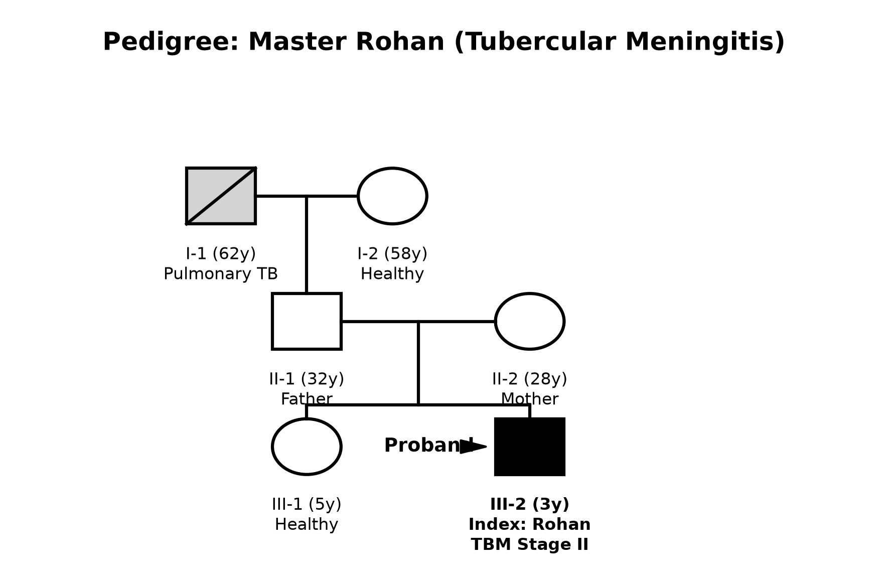
- 2nd-order child born to non-consanguineously married parents.
- Tuberculosis Contact History: Paternal grandfather (residing in the same 2-room household) had chronic productive cough with hemoptysis and was diagnosed with sputum-positive pulmonary tuberculosis 4 months ago; he was treated irregularly with anti-tubercular therapy (ATT).
- Older sister (5 years old) is healthy, thriving, and asymptomatic.
Immunization history
- BCG Status: Mother states the child was born at a primary health centre but BCG vaccination was missed due to local vaccine stock-out.
- No BCG scar visible over the left deltoid on examination.
- Received oral polio vaccine (OPV), Pentavalent (DPT-HepB-Hib 1, 2, 3), and MR 1st dose as per the National Immunization Schedule.
- No optional vaccines (PCV, Varicella, Hepatitis A) were administered.
Dietary history
- The child was exclusively breastfed till 6 months of age; complementary feeding with diluted cow's milk and mashed rice was introduced late at 9 months of age.
- Current intake is significantly compromised due to anorexia, illness, and dysphagia over the past 2 weeks.
| Food Item | Quantity | Calories (kcal) | Protein (g) |
|---|---|---|---|
| Diluted Cow's Milk | 300 mL | 180 | 6.0 |
| Mashed Rice with Dhal | 1 small bowl (~60 g) | 110 | 2.5 |
| Roti (mashed in water) | 1/2 small | 40 | 1.0 |
| Total Daily Intake | 330 | 9.5 |
| 24-Hour Recall | Expected (for Ideal Body Weight ~14 kg) | Observed | Deficit |
|---|---|---|---|
| Calories | 1400 kcal (100 kcal/kg) | 330 kcal | 1070 kcal (76.4% deficit) |
| Protein | 16.8 g (1.2 g/kg) | 9.5 g | 7.3 g (43.5% deficit) |
The expected calories and proteins should be calculated from the ideal body weight, not from current weight.
Socioeconomic and KAP
- Family resides in a semi-pucca, 2-room poorly ventilated house in an urban slum of Varanasi.
- 5 family members live in the household (overcrowding present).
- Paternal grandfather's sputum-positive TB diagnosis was known, but household contact screening and Isoniazid Preventive Therapy (IPT) were not initiated for the children.
- Socioeconomic Status: Lower-middle class according to Modified Kuppuswamy Scale (Score: 8).
Summary of History
"Master Rohan, a 3-year-old male child, 2nd-order offspring of a non-consanguineous marriage from Varanasi, unimmunized with BCG, with a significant household contact history of sputum-positive pulmonary tuberculosis, presented with a 3-week history of subacute low-to-moderate grade fever with evening rise, 10 days of progressive headache and projectile vomiting, 5 days of altered sensorium and excessive irritability, and a recent right-sided focal seizure with right hemiparesis and right convergent strabismus.
In view of the subacute insidious progression, prominent signs of raised intracranial pressure, focal seizure with hemiparesis, and household open-case TB contact, I would like to consider a clinical diagnosis of Chronic Meningoencephalitis, probably Tubercular Meningitis (TBM) - Stage 2 (Intermediate Stage), complicated by Tubercular Vasculitis with Left Middle Cerebral Artery territory infarction and Right CN VI Palsy, with underlying Severe Acute Malnutrition."
General head to toe examination
Pre-Examination Child Behavioral State
- Child State: Child is stuporous, irritable on handling, eyes open intermittently to noxious stimuli, moaning groaning sounds, localized withdrawal to painful stimuli.
- Pediatric Glasgow Coma Scale (pGCS): E3 V3 M4 = 10/15.
Vitals
- Heart Rate: 84 bpm (relative bradycardia for fever, indicating raised intracranial pressure).
- Respiratory Rate: 24/min, regular, no chest retractions.
- Blood Pressure: 108/68 mmHg (>95th percentile for age/height, reflecting Cushing's hemodynamic response).
- Temperature: 38.2°C (100.8°F) axillary.
- Capillary Refill Time: <2 seconds, peripheral pulses well felt.
Anthropometry
| Parameter | Observed | Expected (50th percentile WHO) | Z-Score / Interpretation |
|---|---|---|---|
| Weight | 9.8 kg | 14.3 kg | < -3 SD (Severe Wasting) |
| Length / Height | 88 cm | 96.1 cm | -2 to -3 SD (Moderate Stunting) |
| Weight-for-Height | - | - | < -3 SD (Severe Acute Malnutrition) |
| Head Circumference | 48.2 cm | 49.0 cm | Normal (-1 SD) |
| Mid-Upper Arm Circumference (MUAC) | 11.2 cm | >12.5 cm | < 11.5 cm (SAM) |
Head-to-Toe Markers
- Head & Fontanelle: Anterior fontanelle closed. Macewen's sign ("cracked pot sound") is positive on skull percussion over the frontoparietal junction (indicating dilated ventricles / hydrocephalus).
- BCG Scar: Absent over left deltoid.
- Skin & Hair: Dry, lack-lustre hair with easy pluckability; no erythema nodosum, tuberculides, or petechiae.
- Eyes: No phlyctenular conjunctivitis. Dilated fundoscopic examination reveals:
- Bilateral early papilledema (blurring of nasal disc margins, hyperemic disc, loss of venous pulsations).
- No choroid tubercles identified on direct visualization.
- Lymph Nodes: Multiple discrete, non-tender, non-matted, mobile cervical lymph nodes palpable bilaterally, largest ~1 × 1 cm in the right anterior cervical chain.
- Spine: Normal alignment; no gibbus deformity, tenderness, or cold abscess.
Systemic Examination
CNS Examination
1. Higher Mental Functions & Meningeal Signs
- Level of Consciousness: Drowsy/stuporous (GCS 10/15).
- Meningeal Irritation Signs:
- Neck Rigidity: Markedly positive (marked resistance to passive neck flexion).
- Kernig's Sign: Positive bilaterally at 120° extension.
- Brudzinski's Neck Sign: Positive (reflex flexion of both hips and knees on passive neck flexion).
2. Cranial Nerves
- CN II (Optic): Pupils 3 mm bilaterally, sluggishly reactive to direct and consensual light. Fundus shows bilateral early papilledema.
- CN III, IV, VI: Right eye displays resting inward deviation (esotropia); failure of right eye abduction on horizontal gaze tracking (Right CN VI palsy). Normal vertical gaze and pupillary size.
- CN V (Trigeminal): Corneal reflex present bilaterally. Motor jaw clenching preserved.
- CN VII (Facial): Right-sided Upper Motor Neuron (UMN) facial palsy — flattening of right nasolabial fold, deviation of angle of mouth to left upon crying/grimacing, with preserved forehead wrinkling bilaterally.
- CN VIII: Blinks and turns sluggishly to loud auditory stimuli.
- CN IX, X: Gag reflex present; palate elevates symmetrically; no pooling of secretions in the pharynx.
- CN XI: Symmetrical shoulder movement on noxious stimulation.
- CN XII: Tongue midline in oral cavity; no fasciculations.
3. Motor System Examination
| Motor Parameter | Right Upper Limb | Right Lower Limb | Left Upper Limb | Left Lower Limb |
|---|---|---|---|---|
| Bulk | Mild generalized wasting | Mild generalized wasting | Symmetrical | Symmetrical |
| Tone | Hypertonia (Clasp-knife) | Hypertonia (Clasp-knife) | Normal | Normal |
| Power (MRC Scale) | Grade 2/5 (moves without gravity) | Grade 2/5 | Grade 4+/5 | Grade 4+/5 |
| Biceps Jerk | Exaggerated (3+) | - | Normal (2+) | - |
| Triceps Jerk | Exaggerated (3+) | - | Normal (2+) | - |
| Knee Jerk | - | Exaggerated (3+) | - | Normal (2+) |
| Ankle Jerk | - | Exaggerated (3+) with clonus | - | Normal (2+) |
| Plantar Reflex | Extensor (Babinski +) | Extensor (Babinski +) | Flexor | Flexor |
| Abdominal Reflex | Absent | Absent | Present | Present |
4. Sensory & Cerebellar Examination
- Sensory: Withdraws to painful stimuli in all four limbs (delayed and sluggish in right extremities).
- Cerebellar: Cannot be formally evaluated due to altered sensorium and hemiparesis.
- Involuntary Movements: No choreoathetosis, tremors, or dystonic posturing at rest.
other systems
- Respiratory System: Bilateral symmetrical chest expansion. Vesicular breath sounds heard bilaterally with occasional scattered fine inspiratory crepitations in the right infrascapular region.
- Cardiovascular System: S1, S2 heard normally; no murmurs, rubs, or gallop.
- Abdomen: Soft, non-tender; liver palpable 2 cm below right costal margin (soft, smooth, non-tender span 7 cm); spleen not palpable. No ascites or shifting dullness.
Summary
"Master Rohan, a 3-year-old boy, unimmunized with BCG, with an open household contact of pulmonary tuberculosis, presented with a 3-week history of subacute low-grade fever, headache, projectile vomiting, progressive drowsiness, and a right focal seizure followed by right-sided hemiparesis.
On physical examination, the child is stuporous (GCS 10/15) with bradycardia, relative hypertension, and signs of Severe Acute Malnutrition (MUAC 11.2 cm, Weight-for-Height < -3 SD). Neurological examination confirms marked meningeal irritation (Neck rigidity, Kernig's, Brudzinski's), bilateral papilledema, isolated Right CN VI palsy, Right UMN Facial palsy, and Right-sided Spastic Hemiparesis (Power 2/5, Hypertonia, Hyperreflexia, sustained Ankle Clonus, and Babinski extensor response).
Final Diagnosis: I formulate my diagnosis as Chronic Meningoencephalitis, clinically consistent with Tubercular Meningitis (TBM) - Modified BMRC Stage 2 (Intermediate Stage), complicated by:
- Tubercular Vasculitis with Left MCA Territory Infarction (Internal Capsule) producing Right Spastic Hemiparesis and Right UMN CN VII palsy,
- Right CN VI Palsy (false localizing sign of raised ICP vs basal entrapment),
- Secondary Hydrocephalus / Raised Intracranial Pressure,
- Severe Acute Malnutrition (SAM) without Oedema."
Differential Diagnosis
| Disease Entity | Points in Favor in this Case | Points Against / Differentiating Features |
|---|---|---|
| Tubercular Meningitis (TBM) | Subacute onset (>2 weeks), contact with TB, unimmunized BCG, basal CN VI palsy, focal vasculitic stroke, elevated ICP. | Confirmatory CSF and neuroimaging required. |
| Partially Treated Pyogenic Meningitis | Fever, headache, vomiting, neck rigidity, altered sensorium, focal seizure. | Pyogenic onset is acute/hyperacute (<3–5 days); CSF shows neutrophilic pleocytosis with marked hypoglycorrhachia; responds rapidly to cephalosporins. |
| Viral Encephalitis (e.g., Herpes Simplex) | Acute fever, altered sensorium, focal seizures, focal neurological deficits. | Seizures and coma occur abruptly within 24–48 hours; CSF shows normal or near-normal glucose, lymphocytic pleocytosis, and PCR positivity for HSV/Enterovirus. |
| Fungal Meningitis (Cryptococcal / Histoplasma) | Subacute/chronic course, headache, cranial nerve palsies, raised ICP. | Typically occurs in severely immunocompromised hosts (HIV, primary immunodeficiency, post-transplant); India Ink / Cryptococcal antigen positive in CSF. |
| Brain Abscess (with raised ICP) | Focal seizure, focal hemiparesis, headache, vomiting, papilledema. | Usually preceded by chronic suppurative otitis media (CSOM) or cyanotic congenital heart disease; neuroimaging shows single/multiple ring-enhancing lesions with surrounding vasogenic edema. |
Investigation Protocol & Diagnostic Workup
flowchart TD
    A["Suspected Pediatric TBM"] --> B["1. Urgent Non-Contrast CT / Contrast MRI Brain"]
    B --> C{"Check for Mass Effect / Brainstem Herniation"}
    C -->|No contraindication| D["2. Lumbar Puncture & CSF Analysis"]
    C -->|Severe midline shift / impending herniation| E["Start Empirical ATT + Dexamethasone + Anti-edema therapy; Defer LP"]
    D --> F["CSF Microscopy, Biochemistry, TB-PCR / Xpert MTB/RIF Ultra"]
    A --> G["3. Source & Dissemination Search: Chest X-ray, Gastric Aspirate, Tuberculin Skin Test"]
    A --> H["4. Baseline Organ Profiles: LFT, RFT, Serum Electrolytes (Na+ for SIADH/CSW)"]
1. Cerebrospinal Fluid (CSF) Analysis
- Pressure: Markedly elevated (>200–300 mm H₂O).
- Cytology: Moderately elevated leukocyte count (typically 50–500 cells/µL) with marked lymphocytic predominance (>80%) (neutrophils may appear transiently in early stage 1).
- Biochemistry:
- CSF Protein: Markedly elevated (100–500 mg/dL, and up to >1000 mg/dL in cases with spinal block / Froin's syndrome).
- CSF Glucose: Markedly decreased (<40 mg/dL, with a CSF-to-Blood Glucose Ratio < 0.5, frequently <0.3).
- Cobweb / Pellicle Formation: Classic cobweb clot forming upon standing the CSF tube overnight at 4°C (due to high fibrinogen content).
- Microbiological Confirmation:
- Ziehl-Neelsen / Auramine-Rhodamine Staining: Centrifuged thick smear examination for Acid-Fast Bacilli (AFB).
- CBNAAT / GeneXpert MTB/RIF Ultra on CSF: High sensitivity and specificity; provides rapid detection of Mycobacterium tuberculosis and Rifampicin resistance within 2 hours.
- CSF Mycobacterial Culture (MGIT 960): Gold standard for definitive identification and full drug susceptibility testing (DST).
2. Neuroimaging (Contrast-Enhanced MRI / CT Brain)
- Classic TBM Triad on MRI:
- Basal Meningeal Enhancement: Dense exudates in the interpeduncular cistern, Sylvian fissures, and suprasellar cisterns.
- Hydrocephalus: Symmetrical dilatation of lateral and third ventricles (communicating hydrocephalus due to basal exudates blocking arachnoid villi; or non-communicating due to aqueductal compression by tuberculoma/ependymitis).
- Tubercular Vasculitic Infarctions: Ischemic infarctions characteristically involving the "Tubercular Zone" (basal ganglia, internal capsule, thalamus, and head of caudate nucleus) due to endarteritis of middle cerebral and lenticulostriate perforators.
- Tuberculomas: Ring-enhancing or nodular granulomas with central caseation and surrounding edema.
3. Diagnostic Workup for Primary Focus & Dissemination
- Tuberculin Skin Test (Mantoux): 2 TU PPD intradermally; induration $\ge 10\text{ mm}$ (or $\ge 5\text{ mm}$ in SAM/HIV) read at 48–72 hours is positive. (Note: May be falsely negative in up to 30–50% of advanced TBM due to anergy).
- Chest Radiograph: Anteroposterior view to identify primary complex, mediastinal lymphadenopathy, miliary mottling, or cavitary disease.
- Gastric Aspirate / Induced Sputum for CBNAAT: 3 consecutive morning fasting gastric aspirates for AFB smear and GeneXpert.
- Lymph Node FNAC: If peripheral lymphadenopathy is present, fine needle aspiration for cytology, AFB stain, and CBNAAT.
4. Baseline Monitoring Investigations
- Liver Function Tests (LFT): Baseline SGOT, SGPT, Total/Direct Bilirubin, and Alkaline Phosphatase prior to starting hepatotoxic ATT (Isoniazid, Rifampicin, Pyrazinamide).
- Serum Electrolytes (Na⁺, K⁺): Essential to monitor for hyponatremia secondary to Syndrome of Inappropriate ADH Secretion (SIADH) or Cerebral Salt Wasting (CSW).
- Renal Function Tests & Complete Hemogram: Baseline BUN, Serum Creatinine, Hb, TLC, DLC, ESR.
- HIV Serology: Mandatory in all cases of childhood tuberculosis.
Management Plan
flowchart LR
    A["TBM Treatment Triad"] --> B["1. Anti-Tubercular Therapy (ATT)"]
    A --> C["2. Corticosteroids (Dexamethasone)"]
    A --> D["3. Management of Raised ICP & Hydrocephalus"]
1. Anti-Tubercular Therapy (NTEP 2024 Pediatric Guidelines)
- Regimen: 2 months of 4-drug HRZE followed by 10 months of 3-drug HRE (Total duration: 12 months):
- Intensive Phase (2 Months):
- Isoniazid (H): $10\text{ mg/kg/day}$ (Max: $300\text{ mg/day}$)
- Rifampicin (R): $15\text{ mg/kg/day}$ (Max: $600\text{ mg/day}$)
- Pyrazinamide (Z): $35\text{ mg/kg/day}$ (Max: $1500\text{ mg/day}$)
- Ethambutol (E): $20\text{ mg/kg/day}$ (Max: $1000\text{ mg/day}$)
- Continuation Phase (10 Months):
- Isoniazid (H): $10\text{ mg/kg/day}$
- Rifampicin (R): $15\text{ mg/kg/day}$
- Ethambutol (E): $20\text{ mg/kg/day}$
- Intensive Phase (2 Months):
- Pyridoxine (Vitamin B6) Supplementation: $10\text{–}20\text{ mg/day}$ orally to prevent Isoniazid-induced peripheral neuropathy (especially in SAM).
2. Adjuvant Corticosteroid Therapy
- Indication: Mandatory in all cases of TBM (Stages 1, 2, and 3) to reduce basal exudates, cerebral edema, vasculitis, and incidence of stroke and hydrocephalus.
- Drug & Dosage:
- Intravenous Dexamethasone: $0.4\text{ mg/kg/day}$ (or Oral Prednisolone $2\text{–}4\text{ mg/kg/day}$) for 4 weeks.
- Tapering Schedule: Taper gradually over the next 2 to 4 weeks based on clinical and radiological recovery (Total steroid course: 6–8 weeks).
3. Management of Raised ICP & Hydrocephalus
- Head End Elevation: Nurse the child with head elevated at 30° in the midline to facilitate cerebral venous drainage.
- Medical Management of Cerebral Edema:
- Intravenous 20% Mannitol: $0.5\text{–}1.0\text{ g/kg/dose}$ IV infusion over 20–30 minutes every 6–8 hours for acute ICP spikes (with serum osmolality monitoring).
- Hypertonic Saline (3% NaCl): $3\text{–}5\text{ mL/kg}$ IV bolus for impending herniation.
- Oral Acetazolamide: $20\text{–}40\text{ mg/kg/day}$ in 3 divided doses $\pm$ Oral Furosemide $1\text{ mg/kg/day}$ for communicating, non-progressive hydrocephalus.
- Surgical Management of Hydrocephalus:
- Ventriculoperitoneal (VP) Shunt: Indicated for progressive hydrocephalus, non-communicating hydrocephalus, or failure of medical therapy.
- Endoscopic Third Ventriculostomy (ETV): Considered in selected non-communicating hydrocephalus cases without dense basal scarring.
4. Management of Comorbidities & Complications
- Seizure Control:
- Acute control: IV Midazolam $0.1\text{ mg/kg}$ or IV Lorazepam $0.1\text{ mg/kg}$.
- Maintenance: IV/Oral Levetiracetam ($20\text{–}40\text{ mg/kg/day}$) or Sodium Valproate ($20\text{–}30\text{ mg/kg/day}$).
- Electrolyte Management (SIADH vs CSW):
- SIADH: Euvolemic/Hypervolemic hyponatremia $\rightarrow$ Fluid restriction ($60\text{–}70\%$ maintenance).
- Cerebral Salt Wasting (CSW): Hypovolemic hyponatremia with severe natriuresis $\rightarrow$ Vigorous volume replacement with normal saline $\pm$ Fludrocortisone.
5. Nutritional Rehabilitation & Supportive Care
- Nasogastric Tube Feeding: High-calorie, high-protein feeds (F-75 transitioning to F-100 per WHO SAM protocol).
- Nursing Care for Bedridden Child:
- Two-hourly position changes to prevent decubitus ulcers.
- Methylcellulose eye drops and eye padding for lagophthalmos in facial palsy / comatose state.
- Gentle passive range-of-motion physiotherapy to all four limbs to prevent fixed contractures.
- Long-term Follow-up & Rehabilitation:
- Brainstem Auditory Evoked Response (BERA) at completion of therapy to screen for sensorineural hearing loss.
- Visual acuity, visual evoked potentials (VEP), and serial neurodevelopmental assessment.