
Master Soumyajit, a 3 year old boy, 1st order child born of a non-consanguineous marriage from Kolkata, West Bengal presented with complaints of severe progressive paleness and lethargy noticed since 6 months of age, requiring regular packed red blood cell transfusions every 3 to 4 weeks for the past 2 years, gradual swelling of the abdomen for 1.5 years, and abnormal broadening of facial bones for 1 year.
- Severe, unrelenting pallor and fatigue appearing in late infancy (6 to 12 months of age)
- Transfusion dependence (requiring monthly PRBC transfusions to sustain life)
- Progressive abdominal enlargement (massive splenomegaly and hepatomegaly)
- Abnormal hemolytic facies ("chipmunk" facies from bony marrow expansion)
- Severe growth failure, short stature, and delayed milestones
HOPI
The history is dated back to 6 months of age when the mother first noticed that the child was becoming pale, listless, and uninterested in surroundings.
Always establish the exact age of presentation: Beta-Thalassemia Major does NOT present at birth because protective fetal hemoglobin ($\alpha_2\gamma_2$, HbF) predominates in utero. As the physiological switch from $\gamma$-globin to $\beta$-globin occurs between 3 to 6 months of life, failure of adult hemoglobin ($\alpha_2\beta_2$, HbA) synthesis unmasks severe microcytic hemolytic anemia with profound ineffective erythropoiesis. Meticulously calculate the transfusion and chelation index:
- Age at first transfusion, frequency, volume ($10-15\text{ mL/kg}$), and target pre-transfusion Hb ($9.5-10.5\text{ g/dL}$).
- Total cumulative units transfused ($>10-20\text{ units}$ indicates iron overload).
- Chelation history: Drug name (Deferasirox / Deferiprone / Desferrioxamine), compliance, and side-effect monitoring.
- Onset & Transfusion Dependence:
- Child was seemingly healthy till 6 months of age, when mother noticed progressive pallor of the face, palms, and soles.
- Child became irritable, fatigued quickly during feeds, and stopped gaining weight.
- At 9 months of age, during an episode of viral fever, a local physician documented a Hemoglobin of $4.2\text{ g/dL}$. The child received his first blood transfusion and was diagnosed with Beta-Thalassemia Major.
- Since then, he has been on regular lifelong blood transfusions every 3 to 4 weeks, receiving leukocyte-filtered packed red blood cells. Total transfusions received till date: 26 units.
- Mother notes that towards the end of the 3-week cycle (pre-transfusion), the child becomes pale, breathless on mild exertion, and loses his appetite.
- Abdominal Distension (Hepatosplenomegaly):
- Progressive increase in abdominal girth noticed over the past 1.5 years.
- Mother noticed a hard lump under the left ribs that gradually enlarged downward toward the navel (splenomegaly) Reflects dual pathology: chronic extramedullary hematopoiesis and intense reticuloendothelial clearance of defective, precipitated alpha-chain RBCs.
- Skeletal & Facial Changes (Extramedullary Hematopoiesis):
- Over the past 12 months, parents noticed a striking change in the child's facial appearance.
- Forehead became prominent and bulging, cheekbones became unusually elevated, and the bridge of the nose appeared flattened (chipmunk facies) Massive compensatory expansion of the erythroid marrow space (up to 20-30 times normal) within the skull and facial bones causes diploic space widening and cortical bone thinning.
- Iron Overload & Chelation History:
- Child has received ~26 transfusions ($>120\text{ mL/kg}$ cumulative PRBC).
- Serum Ferritin measured 2 months ago was $1,850\text{ ng/mL}$ (elevated; threshold to start chelation is $>1,000\text{ ng/mL}$).
- Started on oral Deferasirox ($20\text{ mg/kg/day}$) 4 months ago; parents report irregular compliance due to gastrointestinal upset.
- Negative History:
- No history of cola-colored or black urine Excludes acute intravascular hemolysis / hemoglobinuria.
- No history of severe bone pain, dactylitis (swelling of hands/feet), or acute chest pain Rules out vaso-occlusive crisis of Sickle Cell Anemia.
- No history of sudden deepening of pallor with high fever Rules out Parvovirus B19 aplastic crisis.
- No history of transfusion reactions (urticaria, chills, rigors, dyspnea) during transfusions.
- No history of bleeding from gums, petechiae, or purpuric spots.
Past History
- Diagnosed with Beta-Thalassemia Major at 9 months of age via Hemoglobin HPLC.
- Transfused every 3-4 weeks since age 9 months.
- No history of splenectomy or cholecystectomy.
Family history
- Born of a non-consanguineous marriage.
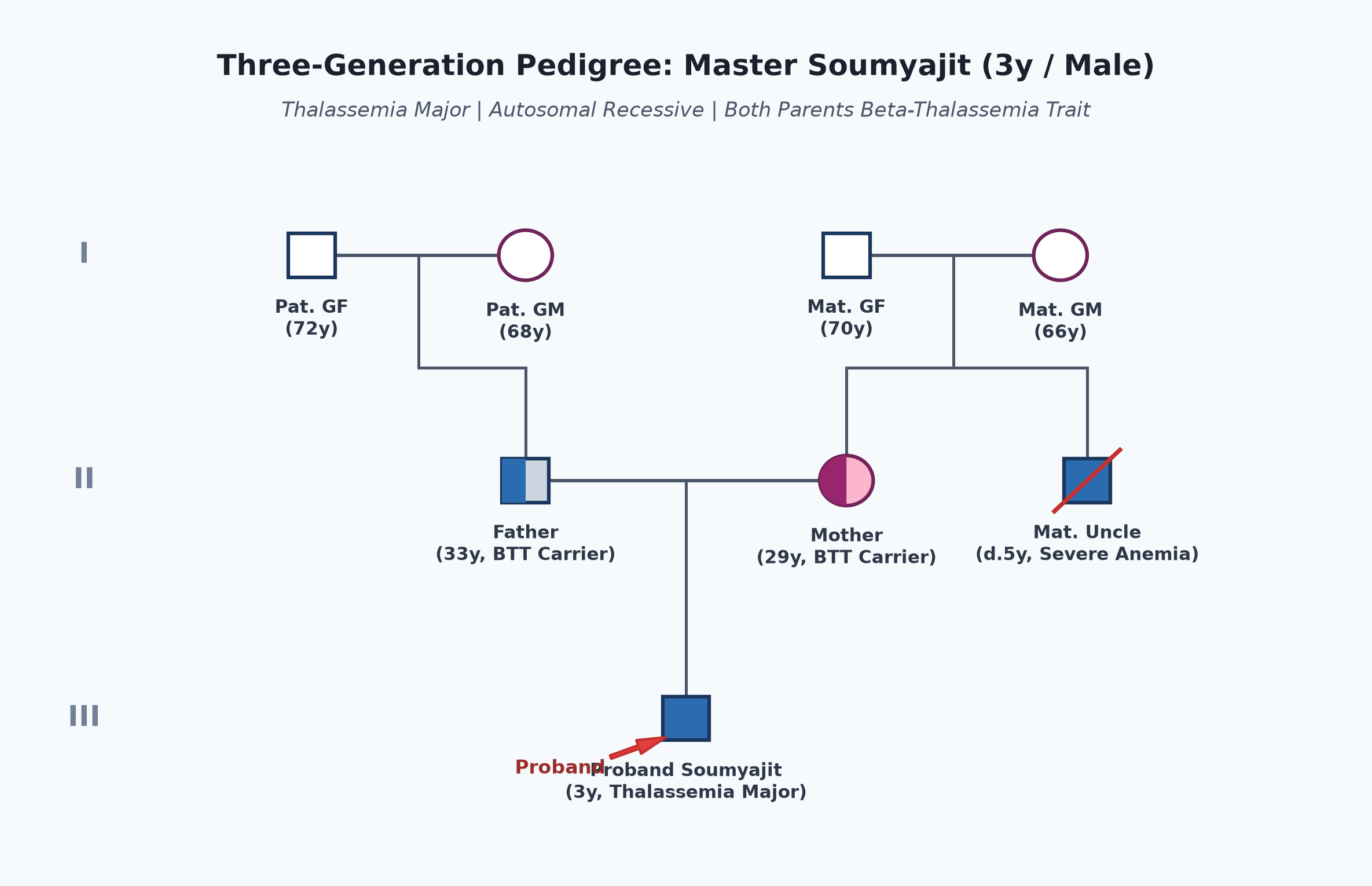
- Father 33 years, office clerk; mother 29 years, homemaker; both are confirmed carriers of Beta-Thalassemia Trait (Minor).
- Family Lineage: Maternal uncle suffered from severe childhood anemia and died at 5 years of age (undiagnosed thalassemia major).
- Proband is an only child; parents received antenatal counseling for future pregnancies.
Immunization history
- Fully immunized as per the National Immunization Schedule (UIP) up to 3 years.
- Additional vaccines: Received Hepatitis B vaccine (full 3-dose course + booster) and Hepatitis A vaccine (mandatory prior to frequent transfusions).
- Pneumococcal and Meningococcal vaccines planned in view of future splenectomy risk.
Dietary history
- Consumes a soft home diet consisting of rice, dal, and vegetables.
- Parents strictly avoid iron-rich foods (liver, red meat, green leafy vegetables) and iron-fortified cereals.
- Mother gives black tea with meals (tannins inhibit intestinal non-heme iron absorption).
| Food Item | Quantity | Calories (kcal) | Protein (g) |
|---|---|---|---|
| Boiled Rice | 120 g | 156 | 3.2 |
| Moong Dal | 1 katori | 90 | 5.5 |
| Cow's Milk (toned) | 250 mL | 150 | 8.0 |
| Mashed Vegetables | 1 cup | 70 | 1.4 |
| Total Observed Daily Intake | — | 466 kcal | 18.1 g |
24-Hour Recall Deficit Analysis
$$ \text{Ideal Body Weight (IBW for 3 years, 50th centile WHO)} = 14.3\text{ kg} $$| Nutrient | Expected Intake (ICMR-NIN 2024 for IBW 14.3 kg) | Observed Intake | Deficit | Percentage Deficit |
|---|---|---|---|---|
| Energy (kcal) | $14.3\text{ kg} \times 80\text{ kcal/kg} = 1144\text{ kcal}$ | 466 kcal | 678 kcal | 59.3% Deficit |
| Protein (g) | $14.3\text{ kg} \times 1.1\text{ g/kg} = 15.7\text{ g}$ | 18.1 g | Nil (Adequate) | 0% Deficit |
The expected calories and proteins should be calculated from the ideal body weight, not from current weight.
Socioeconomic and KAP
- Belongs to Modified BG Prasad Socioeconomic Class III (Middle Class).
- Urban residence in Kolkata with ready access to a thalassemia transfusion center.
- Parents understand that transfusions sustain life, but are financially stressed by chelation costs and anxious regarding curative stem cell transplantation.
Summary of History
Master Soumyajit, a 3-year-old boy, 1st order child born of non-consanguineous parents who are both beta-thalassemia trait carriers from Kolkata, presented with severe progressive pallor since 6 months of age, transfusion dependence requiring 3-4 weekly PRBC transfusions for 2 years (total 26 units), progressive splenomegaly, hemolytic bone changes, and elevated ferritin ($1,850\text{ ng/mL}$), without features of sickling vaso-occlusive crisis, aplastic crisis, or transfusion reactions.
I would like to think of a hereditary hemolytic anemia secondary to Beta-Thalassemia Major, transfusion-dependent, complicated by extramedullary hematopoiesis (splenomegaly, hemolytic facies), secondary hemosiderosis, and severe failure to thrive.
General head to toe examination
- Child Behavioral State: Quiet wakefulness (Prechtl State 3), listless, cooperative.
- Vitals:
- Pulse Rate: 118 beats/minute (resting tachycardia), regular, bounding / water-hammer character.
- Respiratory Rate: 26 breaths/minute, regular, abdominothoracic.
- Blood Pressure: $98/52\text{ mmHg}$ (wide pulse pressure $\approx 46\text{ mmHg}$ indicating a hyperdynamic circulatory state).
- Temperature: $36.8^\circ\text{C}$ (afebrile).
- Anthropometry:
| Parameter | Observed | Expected (50th WHO) | Z-score / Centile | Inference |
|---|---|---|---|---|
| Weight | 11.0 kg | 14.3 kg | $< -2\text{ SD}$ | Moderate Underweight |
| Height | 87 cm | 96 cm | $< -2\text{ SD}$ | Moderate Stunting |
| Head Circumference | 49.0 cm | 49.5 cm | Normal | Normal head growth |
- Craniofacial & Hemolytic Facies:
- Classical "Chipmunk / Thalassemic Facies":
- Frontal bossing and prominent parietal eminences.
- Depressed, flattened nasal bridge.
- Prominent malar eminences (malar prominence).
- Maxillary hyperplasia with upward retraction of the upper lip exposing protruding upper incisors and dental malocclusion.
- Classical "Chipmunk / Thalassemic Facies":
- General Findings:
- Pallor: Severe pallor in conjunctiva, mucosal surfaces, palms, and nail beds.
- Icterus: Mild lemon-yellow icterus in the superior bulbar conjunctiva (unconjugated hyperbilirubinemia from chronic extravascular hemolysis).
- Skin Pigmentation: Generalized grayish-bronze / muddy skin discoloration (cutaneous hemosiderosis / melanin stimulation).
- Lymphadenopathy / Edema / Cyanosis: Absent.
Systemic Examination
Abdomen
- Inspection:
- Protuberant, distended abdomen with prominent superficial non-tortuous veins.
- Umbilicus central, stretched; no caput medusae.
- Palpation:
- Soft, non-tender throughout; no guarding.
- Spleen:
- Massive Splenomegaly: Palpable 6.0 cm below the left costal margin along its long axis toward the right iliac fossa.
- Firm consistency, smooth surface, prominent splenic notch palpable on anterior border, non-tender, no friction rub.
- Liver:
- Hepatomegaly: Palpable 4.0 cm below right costal margin in midclavicular line.
- Firm in consistency, smooth surface, rounded edge, non-tender; total Liver Span is 11.0 cm (normal $\approx 7.5-8\text{ cm}$).
- Kidneys: Not palpable.
- Percussion: Dull over organomegaly; central tympany; no shifting dullness (no ascites).
- Auscultation: Normal bowel sounds; no venous hum or bruits.
Cardiovascular System (CVS)
- Precordium hyperdynamic; visible apex beat in 5th intercostal space outside the midclavicular line.
- Normal $S_1$ and $S_2$; Grade 3/6 ejection systolic hemic murmur heard best over the pulmonary area and left sternal border (turbulent flow across normal valves from severe chronic anemia).
- No gallop rhythm, JVP not elevated.
Respiratory & Central Nervous Systems
- Lungs clear bilaterally with normal vesicular breath sounds.
- CNS completely intact; alert, responsive; normal tone, power (5/5), and reflexes.
other systems
- Musculoskeletal: No active bone tenderness; extremities thin.
Summary
Master Soumyajit, a 3-year-old boy, born of non-consanguineous parents who are both beta-thalassemia carriers from Kolkata, presented with a 2-year history of transfusion dependence requiring monthly PRBC transfusions, progressive hepatosplenomegaly, and facial changes. Physical examination confirms severe pallor, lemon-yellow icterus, bronze hyperpigmentation, thalassemic chipmunk facies, hyperdynamic circulation with wide pulse pressure and a hemic murmur, marked splenomegaly (6 cm), and hepatomegaly (liver span 11 cm), with an elevated ferritin of $1,850\text{ ng/mL}$.
The clinical presentation is pathognomonic for Transfusion-Dependent Beta-Thalassemia Major, complicated by extramedullary hematopoiesis, hyperdynamic circulation, secondary hemosiderosis, and growth faltering.
Differential Diagnosis
| Disease | Points IN FAVOR | Points AGAINST |
|---|---|---|
| Beta-Thalassemia Major | Onset in late infancy (6-12m), lifelong transfusion dependence, chipmunk facies, massive splenomegaly, HPLC confirms HbF >80% | Primary Diagnosis |
| HbE-Beta Thalassemia | Common in West Bengal/Assam, severe anemia, hepatosplenomegaly, hemolytic facies | HPLC demonstrates prominent HbE peak along with HbF; clinically ranges from mild non-transfusion dependent to severe |
| Sickle-Beta Thalassemia | Microcytic anemia, splenomegaly, consanguinity | Vaso-occlusive bone crises and dactylitis are absent; HPLC shows HbS peak; sickle solubility test negative |
| Hereditary Spherocytosis | Hemolytic anemia, jaundice, splenomegaly, gallstones | Autosomal dominant usually, spherocytes on blood smear, normal HPLC with elevated MCHC, negative HbF elevation |
| Autoimmune Hemolytic Anemia (AIHA) | Acquired severe pallor, icterus, splenomegaly | Sudden onset, lacks chipmunk facies, no microcytosis, Direct Antiglobulin Test (Coombs) is positive |
Investigation Protocol & Diagnostic Workup
flowchart TD
    A["Child with Chronic Microcytic Hemolytic Anemia & Hepatosplenomegaly"] --> B["CBC, Peripheral Smear & Reticulocyte Count"]
    B --> C["Identify Severe Microcytic Hypochromic Anemia with Nucleated RBCs"]
    C --> D["Hemoglobin HPLC / Capillary Zone Electrophoresis"]
    D --> E{"HbF > 70-90% & Virtually Absent HbA?"}
    E -->|Yes| F["Confirm Beta-Thalassemia Major; Screen Both Parents (HbA2 > 3.5%)"]
    F --> G["Iron Overload Assessment: Serum Ferritin, Liver Function Tests"]
    G --> H["Imaging: Skull Radiograph (Hair-on-End Sign) & Abdominal USG"]
    H --> I["Pre-Transfusion Viral Screening: NAT for HIV, HBV, HCV"]
    I --> J["Initiate Hypertransfusion Regimen & Oral Iron Chelation (Deferasirox)"]
1. Confirmatory Hemoglobin Analysis (HPLC)
- High-Performance Liquid Chromatography (Bio-Rad Variant II):
- HbF (Fetal Hemoglobin): Markedly elevated at $88.5\%$ (diagnostic hallmark).
- HbA (Adult Hemoglobin): Virtually absent ($<2.0\%$) in $\beta^0/\beta^0$ or markedly reduced ($<10\%$) in $\beta^0/\beta^+$.
- HbA2: Variable ($1.8-3.2\%$).
- Parental HPLC Screening:
- Both father and mother demonstrate elevated $\text{HbA}_2 > 3.5\%$ (Father $5.2\%$, Mother $4.8\%$), confirming that both parents are heterozygous carriers of Beta-Thalassemia Minor.
2. Complete Blood Count & Blood Smear
- Hemoglobin (Pre-transfusion): $6.2\text{ g/dL}$.
- RBC Indices: Severe microcytosis (MCV $61.5\text{ fL}$, normal $75-87\text{ fL}$), severe hypochromia (MCH $18.2\text{ pg}$, normal $24-30\text{ pg}$), RDW markedly elevated ($26.4\%$).
- Peripheral Blood Smear: Marked anisopoikilocytosis, microcytes, hypochromia, numerous target cells (leptocytes), tear-drop cells, basophilic stippling, and numerous nucleated RBCs (erythroblasts: 38 per 100 WBCs) reflecting intense extramedullary erythropoiesis.
- Reticulocyte Count: Corrected Reticulocyte Count $3.8\%$ (elevated, but inappropriately low for the profound degree of anemia due to massive intramedullary apoptosis of erythroid precursors).
3. Iron Overload Studies
- Serum Ferritin: $1,850\text{ ng/mL}$ (target in well-chelated children is $<1,000\text{ ng/mL}$).
- Serum Iron: $210\text{ mcg/dL}$ (elevated); TIBC: $240\text{ mcg/dL}$ (decreased); Transferrin Saturation: $87.5\%$ (markedly elevated).
4. Radiological Imaging
- Skull Radiograph (Lateral View):
- Classical "Hair-on-End" (Crew-cut) appearance: widening of the diploic space with thinning of the outer table and fine vertical striations of bone trabeculae perpendicular to the tables.
- Abdominal Ultrasound: Confirms marked homogeneous hepatosplenomegaly; no gallstones or biliary sludge visualized.
Management Plan
1. Hypertransfusion Protocol (Standard of Care)
- Target Hemoglobin: Maintain pre-transfusion Hemoglobin strictly between $9.5$ and $10.5\text{ g/dL}$ (post-transfusion Hb $13.5-14.0\text{ g/dL}$).
- Clinical Rationale: Maintaining pre-transfusion $\text{Hb} > 9.5\text{ g/dL}$ completely suppresses endogenous ineffective erythropoiesis, prevents skeletal facial deformities, halts progressive hepatosplenomegaly, prevents cardiomegaly, and supports normal somatic growth.
- Transfusion Specification:
- Transfuse Leukocyte-Depleted (leukofiltered) Packed Red Blood Cells (PRBC), crossmatch-compatible, phenotype-matched for Rh and Kell antigens, screened by Nucleic Acid Testing (NAT) for HIV, HBV, and HCV.
- Dose: $10-15\text{ mL/kg}$ per transfusion, infused over 3–4 hours every 3 to 4 weeks.
2. Iron Chelation Therapy
- Initiation Criteria: Initiated when Serum Ferritin $>1,000\text{ ng/mL}$ OR after receiving $>10-20$ transfusions (typically around 2–3 years of age).
- First-Line Oral Chelator:
- Deferasirox (DFX): Administer once daily at $20-30\text{ mg/kg/day}$ (as dispersible tablets dissolved in water/apple juice on an empty stomach 30 minutes before food).
- Monitoring: Monthly serum creatinine and urine protein-to-creatinine ratio (renal tubular toxicity), monthly ALT/AST, and quarterly serum ferritin.
- Alternative / Combination Chelators (for Severe Iron Overload):
- Deferiprone (DFP): Oral $75-100\text{ mg/kg/day}$ divided tid; highly effective for cardiac iron removal; requires weekly Absolute Neutrophil Count (ANC) monitoring due to the risk of agranulocytosis.
- Deferoxamine (DFO): Subcutaneous infusion $30-50\text{ mg/kg/day}$ over 8–12 hours using a portable infusion pump 5–7 nights/week.
3. Adjuvant Medical & Nutritional Therapy
- Folic Acid: Daily oral supplementation ($1-2\text{ mg/day}$) to sustain active erythropoiesis.
- Strict Avoidance of Iron: Prohibit all iron-containing tonics, vitamin syrups with iron, and iron-fortified baby foods.
- Dietary Advice: Encourage drinking black tea with meals (tea polyphenols bind dietary iron).
4. Definitive Curative Therapy
- Allogeneic Hematopoietic Stem Cell Transplantation (HSCT):
- The only widely established curative modality.
- Best outcomes achieved with an HLA-identical matched sibling donor (MSD) prior to the onset of severe iron overload and hepatomegaly (Pesaro Risk Class 1: disease-free survival $>90\%$).
- Novel Gene Therapy: Autologous CD34+ cells transduced with lentiviral vector expressing $\beta$-globin (Betibeglogene autotemcel) or CRISPR-Cas9 gene editing (Exagamglogene autotemcel / Casgevy).
5. Indications for Splenectomy (Deferred Ideally Until >5-6 Years)
- Reserved strictly for children with annual PRBC transfusion requirements exceeding $>200-220\text{ mL/kg/year}$, symptomatic hypersplenism (worsening leukopenia/thrombocytopenia), or massive mechanical discomfort.
- Mandatory pre-splenectomy immunization against Streptococcus pneumoniae (PCV13 + PPSV23), Neisseria meningitidis (MenACWY), and Haemophilus influenzae type b (Hib) at least 4 weeks prior, followed by lifelong daily oral Penicillin V prophylaxis.