
Master Aarav, a 6-month-old male infant, 1st order child of non-consanguineous marriage from Patna, Bihar presented with complaints of progressive enlargement of head size since 2 months of age, inability to hold his neck, and downward rolling of the eyes noticed over the past 2 months.
- Rapid, progressive enlargement of the head (macrocephaly)
- Tense, bulging anterior fontanelle with splayed cranial sutures
- Downward gaze preference / sclera visible above iris (Setting Sun phenomenon)
- Failure to achieve neck holding due to disproportionate head weight
- Associated cystic spinal swelling (meningomyelocele) or lower limb paralysis
HOPI
- Progressive Head Enlargement:
- The infant was born with a head circumference of 35 cm (50th percentile) and had an uneventful early neonatal course after undergoing surgical repair of a lumbosacral spinal defect on Day 3 of life.
- Around 2 months of age, the mother noticed that the infant's head began growing unusually fast; caps and bonnets became tight within days.
- The calvarium became globular and disproportionately large compared to his small facial features (craniofacial disproportion).
- Fontanelle & Scalp Changes:
- The mother observed that the soft spot on top of the head (anterior fontanelle) was constantly swollen, tense, and bulging, even when the baby was calm, asleep, or held upright.
- Prominent, bluish, engorged blood vessels appeared over his forehead and scalp, becoming particularly visible when the infant cried.
- The infant exhibited frequent bouts of high-pitched irritable crying, poor feeding, and occasional effortless vomiting.
- Eye Signs (Setting Sun Phenomenon):
- Over the past 6 weeks, parents noticed that both eyes tended to deviate downwards, such that a wide white rim of sclera was constantly visible between the upper eyelid and the colored iris ("like a setting sun").
- Motor Development & Spine:
- At 6 months of age, the infant is unable to support or balance his head when pulled into a sitting position (complete head lag due to mechanical head heaviness).
- Social smile and cooing are present; he fixates on the mother's face.
- The infant has decreased spontaneous movement of both lower limbs with inward turning of both feet (bilateral clubfoot / talipes equinovarus) noticed since birth.
- Urine dribbles continuously in small drops throughout the day without a forceful continuous stream.
Pertinent Negatives in HOPI:
- No history of fever, neck stiffness, altered sensorium, or purulent ear discharge (rules out acute post-meningitic ventriculitis).
- No history of stridor, hoarse cry, choking during feeding, or apnoeic spells (rules out acute symptomatic brainstem compression from Arnold-Chiari Type II malformation).
- No history of loss of previously acquired milestones (rules out neurodegenerative megalencephalies like Alexander or Canavan disease).
- No history of infantile spasms or hypopigmented skin patches (rules out Tuberous Sclerosis with subependymal giant cell astrocytoma).
- No history of family members with large heads or adducted thumbs (rules out X-linked aqueductal stenosis / Bickers-Adams syndrome).
Past History
- Born with a ruptured lumbosacral cystic swelling (Meningomyelocele, $4\times 4\text{ cm}$), which underwent primary surgical excision and neural placode closure on Day 3 of life at a tertiary centre. Post-operative recovery was uneventful.
- No history of neonatal sepsis, meningitis, or intracranial hemorrhage.
Family history
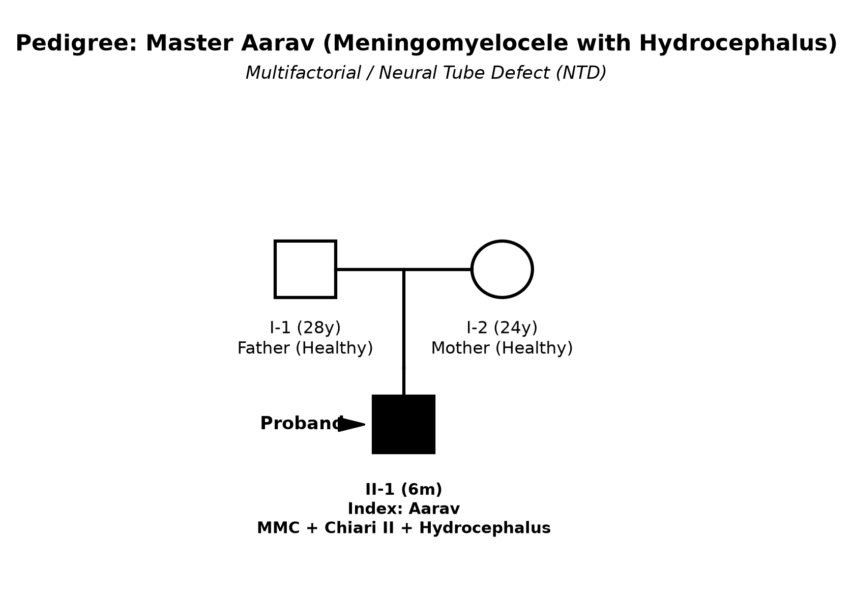
- 1st-order child born to non-consanguineously married parents.
- Mother did not receive periconceptional folic acid supplementation during the first trimester.
- No family history of neural tube defects, hydrocephalus, or unexplained infant deaths.
Immunization history
- Immunized up to age with BCG, OPV 0-3, Pentavalent 1-3, Rota 1-3, and fIPV 1-2 as per the National Immunization Schedule.
- BCG scar is present on the left deltoid.
Dietary history
- Exclusively breastfed on demand (8–10 times per day); latches well; complementary feeding not yet initiated.
| Food Item | Quantity | Calories (kcal) | Protein (g) |
|---|---|---|---|
| Exclusive Breast Milk | ~650 mL/day | 435 | 7.8 |
| Total Daily Intake | 435 | 7.8 |
| 24-Hour Recall | Expected (for Ideal Body Weight ~7.5 kg) | Observed | Deficit |
|---|---|---|---|
| Calories | 750 kcal (~100 kcal/kg) | 435 kcal | 315 kcal (42.0% deficit) |
| Protein | 9.0 g (1.2 g/kg) | 7.8 g | 1.2 g (13.3% deficit) |
The expected calories and proteins should be calculated from the ideal body weight, not from current weight.
Socioeconomic and KAP
- Family resides in a semi-pucca house in suburban Patna.
- Father is a daily-wage technician; Mother is a homemaker.
- Socioeconomic Status: Lower-middle class (Modified Kuppuswamy Score: 10).
- Parents understand the need for surgical cerebrospinal fluid diversion (VP shunt) and long-term bladder management.
Summary of History
"Master Aarav, a 6-month-old male infant, 1st-order child of non-consanguineous parents, with a surgically repaired lumbosacral meningomyelocele on Day 3 of life, presented with progressive head enlargement since 2 months of age, tense bulging anterior fontanelle, dilated scalp veins, setting sun eye phenomenon, and head lag.
The infant has associated lower motor neuron paraplegia with bilateral clubfoot and continuous urinary dribbling (neurogenic bladder), with preserved social and language milestones, and with No history of stridor, apnea, or acute ventriculitis.
In view of the post-meningomyelocele status, rapid head expansion exceeding WHO percentiles, and typical semiological ocular signs, I would like to consider a clinical diagnosis of Congenital Hydrocephalus, secondary to Arnold-Chiari Malformation Type II associated with Repaired Lumbosacral Meningomyelocele, currently with Compensated Raised Intracranial Pressure, with L5–S1 Lower Motor Neuron Paraplegia and Neurogenic Bladder."
General head to toe examination
Pre-Examination Child Behavioral State
- Child State: Infant is calm, lying supine in quiet wakefulness, non-crying (Prechtl State 3).
Vitals
- Pulse Rate: 118 bpm, regular, normal volume.
- Respiratory Rate: 28/min, regular, no stridor or chest indrawing.
- Blood Pressure: 86/54 mmHg (normal for age).
- Temperature: 37.0°C (98.6°F), afebrile.
Cranial & Skull Examination (Detailed Semiology)
| Parameter | Observed Value | Expected (50th percentile WHO) | Interpretation |
|---|---|---|---|
| Head Circumference (HC) | 48.5 cm | 43.3 cm | Macrocephaly ($>+3\text{ SD}$ above mean) |
| Weight | 6.8 kg | 7.9 kg | Underweight (-1.5 SD) |
| Length | 65.0 cm | 67.6 cm | Normal (-1 SD) |
- Cranial Inspection:
- Craniofacial Disproportion: Massive, globular, rounded calvarium towering over a small, triangular facies.
- Frontal Bossing: Prominent overhanging forehead with depressed nasal root.
- Dilated Scalp Veins: Prominent, engorged collateral scalp veins radiating over the frontal and temporal scalp.
- Scalp skin is thin, stretched, translucent, and shiny.
- Fontanelles & Sutures:
- Anterior Fontanelle: $5.0 \times 5.0\text{ cm}$, markedly tense, bulging above the bone margins, non-pulsatile in the upright sitting position.
- Sutures: Coronal, sagittal, and lambdoid sutures are widely separated ($>1.0\text{ cm}$ diastasis on palpation).
- Percussion & Special Tests:
- Macewen's Sign ("Cracked Pot Sound"): Percussion over the frontoparietal suture line elicits a resonant, high-pitched cracked pot sound.
- Transillumination Test: Symmetrical light glow $<2.0\text{ cm}$ (indicates presence of remaining cerebral cortical mantle).
- Ocular Signs:
- Setting Sun Sign (Sunset Phenomenon): Present bilaterally at rest; both globes deviated downwards exposing the upper sclera.
- Pupillary Reflexes: 2.5 mm bilaterally, briskly reactive to light.
- Fundoscopy: Pale optic disc with sharp margins (early secondary optic atrophy); no acute flaming papilledema.
Spine & Perineal Examination
- Spine: Well-healed, transverse $5\text{ cm}$ surgical scar in the lumbosacral region (L4–S1 level); no CSF leak, redness, fluctuant collection, or sinus tract.
- Perineum & Sphincters:
- Patulous, lax anal sphincter with gaping anal orifice.
- Anal Wink Reflex: Absent (pinprick around perianal S4–S5 dermatome fails to elicit reflex anal contraction).
- Palpable distended bladder up to the umbilicus; gentle suprapubic pressure elicits expression of urine (overflow incontinence / lower motor neuron neurogenic bladder).
- Extremities: Symmetrical bilateral Talipes Equinovarus (Clubfoot) deformities (fixed ankle equinus, heel varus, forefoot adduction).
Systemic Examination
CNS Examination
1. Higher Mental Functions & Cranial Nerves
- Consciousness: Alert, responsive to visual stimuli; smiles and coos socially.
- Cranial Nerves:
- CN II: Fixates and tracks bright objects within visual field; setting sun sign present.
- CN III, IV, VI: Extraocular movements full except for limited spontaneous upward gaze.
- CN VII: Symmetrical smile and facial movements; no facial palsy.
- CN IX, X, XII: Good sucking and swallowing; normal cry; no stridor or tongue fasciculations.
2. Motor System Examination
| Motor Parameter | Upper Limbs | Lower Limbs |
|---|---|---|
| Spontaneous Movements | Normal, active reaching | Severely reduced, minimal spontaneous kicks |
| Muscle Bulk | Normal for age | Marked wasting of thigh and calf muscles |
| Tone | Normal | Hypotonia / Flaccidity |
| Power (MRC Scale) | Grade 5/5 (moves actively against gravity) | Grade 1–2/5 (Hip flexors 2/5, Knee extensors 1/5, Ankle dorsiflexors 0/5) |
| Biceps / Triceps Jerk | Normal (2+) | - |
| Knee Jerk | - | Absent (0) bilaterally |
| Ankle Jerk | - | Absent (0) bilaterally |
| Plantar Reflex | Flexor | No response (Mute plantars) |
3. Sensory System
- Pain sensation (pinprick withdrawal): Intact in upper limbs and trunk down to the L2 dermatome; absent / anaesthetic below the L4 dermatome (saddle area, legs, and feet).
other systems
- Respiratory System: Vesicular breath sounds bilaterally; clear chest; no stridor or wheeze.
- Cardiovascular System: Heart sounds S1, S2 heard normally; no murmurs.
- Abdomen: Soft; distended urinary bladder palpable; liver 1.5 cm below costal margin (soft, smooth); spleen not palpable.
Summary
"Master Aarav, a 6-month-old male infant, born with a lumbosacral meningomyelocele surgically repaired on Day 3 of life, presented with progressive macrocephaly since 2 months of age, inability to hold his neck, setting sun eye phenomenon, and continuous urinary dribbling.
Physical examination reveals a head circumference of 48.5 cm ($>+3\text{ SD}$), craniofacial disproportion, tense bulging anterior fontanelle ($5\times 5\text{ cm}$), suture diastasis, dilated scalp veins, positive Macewen's sign, and bilateral setting-sun sign. Systemic examination reveals a well-healed lumbosacral scar, bilateral lower motor neuron flaccid paraplegia below L4, absent deep tendon reflexes, absent anal wink, neurogenic bladder with overflow incontinence, and bilateral talipes equinovarus.
Final Diagnosis: I formulate my diagnosis as Congenital Hydrocephalus, secondary to Arnold-Chiari Malformation Type II associated with Repaired Lumbosacral Meningomyelocele, with L4–S1 Lower Motor Neuron Flaccid Paraplegia and Neurogenic Bladder, and Bilateral Talipes Equinovarus, currently with Compensated Raised Intracranial Pressure and No clinical signs of active shunt infection or ascending ventriculitis."
Differential Diagnosis
| Disease Entity | Points in Favor in this Case | Points Against / Differentiating Features |
|---|---|---|
| Congenital Hydrocephalus (Chiari II + MMC) | Lumbosacral MMC repair, rapid macrocephaly ($>+3\text{ SD}$), tense fontanelle, sunset sign, dilated scalp veins, flaccid paraplegia. | Classic presentation; confirmed on neuroimaging showing tectal beaking, cerebellar tonsillar descent, and marked ventriculomegaly. |
| Congenital Aqueductal Stenosis (Isolated) | Early progressive macrocephaly, bulging fontanelle, setting sun sign. | Aqueductal stenosis occurs in the absence of spinal dysraphism/MMC; lower limbs show pyramidal spastic paraparesis rather than LMN flaccid paralysis. |
| Dandy-Walker Malformation | Progressive hydrocephalus, bulging fontanelle, developmental delay. | Characterized by cystic enlargement of the posterior fossa, prominent occiput, and agenesis/hypoplasia of the cerebellar vermis on MRI. |
| Post-Meningitic / Post-Hemorrhagic Hydrocephalus | Communicating hydrocephalus, suture splaying, raised ICP signs. | History of documented bacterial meningitis, prolonged fever, or prematurity with intraventricular hemorrhage; arachnoid scarring on MRI. |
| Megalencephaly (Alexander / Canavan Disease) | Macrocephaly, head lag, delayed gross motor milestones. | Megalencephaly is characterized by progressive neuroregression, early seizures, absence of suture diastasis/sunset sign, and MRI showing diffuse white matter leukodystrophy without ventricular enlargement. |
Investigation Protocol & Diagnostic Workup
flowchart TD
    A["Suspected Hydrocephalus with MMC"] --> B["1. Neurosonogram (Transfontanellar USG)"]
    B --> C["Dilated Lateral & Third Ventricles (Evans Index > 0.3)"]
    C --> D["2. MRI Brain & Whole Spine (with CISS / FIESTA sequences)"]
    D --> E["Chiari II: Tectal Beaking, Tonsillar Herniation, Hydrocephalus, Tethered Cord"]
    A --> F["3. Renal & Bladder Ultrasound (Hydronephrosis, Trabeculated Bladder)"]
    A --> G["4. Urodynamic Studies (Detrusor-Sphincter Dyssynergia / High-Pressure Bladder)"]
1. Neuroimaging (First-Line Diagnostic Standard)
- Transfontanellar Neurosonography (NSG):
- Non-invasive bedside screening through the open anterior fontanelle.
- Measures Anterior Horn Width (AHW), Thalamo-Occipital Distance (TOD), and Evans Index: $$\text{Evans Index} = \frac{\text{Maximum width of frontal horns}}{\text{Maximum internal diameter of skull}}$$ An Evans Index $>0.30$ establishes significant ventriculomegaly.
- MRI Brain and Whole Spine with 3D CISS / FIESTA Sequences:
- Brain Findings (Chiari II Malformation): Beaking of the midbrain tectum, downward herniation of the cerebellar vermis and fourth ventricle through the foramen magnum, hypoplastic tentorium, agenesis/thinning of corpus callosum, and severe lateral and third ventricular dilatation.
- Spine Findings: Level of placode closure, low-lying conus medullaris (tethered cord below L2), syrinx / hydromyelia.
2. Urological & Renal Evaluation
- Ultrasound Kidneys, Ureters, and Bladder (KUB): Essential baseline screening for bilateral hydroureteronephrosis, cortical thinning, post-void residual volume, and thickened trabeculated bladder wall.
- Micturating Cystourethrogram (MCU / VCUG): Identifies Vesicoureteral Reflux (VUR, Grades I–V) and bladder neck incompetence.
- Urodynamic Studies (UDS): Quantifies detrusor leak point pressure (DLPP $>40\text{ cm H}_2\text{O}$ carries high risk of upper tract renal deterioration).
Management Plan
flowchart LR
    A["Multidisciplinary MMC & Hydrocephalus Care"] --> B["1. Neurosurgical CSF Diversion (VP Shunt / ETV)"]
    A --> C["2. Urological Management (CIC & Anticholinergics)"]
    A --> D["3. Orthopedic Care & Clubfoot Splinting"]
    A --> E["4. Recurrence Prevention (Folic Acid)"]
1. Neurosurgical Management of Hydrocephalus
- Ventriculoperitoneal (VP) Shunt Surgery (Gold Standard):
- Placement of a silicone catheter system with a unidirectional pressure-regulating valve connecting the right frontal (Kocher's point) or parietal horn to the peritoneal cavity.
- Selection of Valve: Medium-pressure or programmable differential-pressure valve with anti-siphon device.
- Endoscopic Third Ventriculostomy with Choroid Plexus Cauterization (ETV/CPC):
- Alternative surgical procedure in selected centers for non-communicating hydrocephalus to avoid permanent prosthetic shunt dependency.
2. Urological Management (Preservation of Renal Function)
- Clean Intermittent Catheterization (CIC):
- Initiate sterile/clean catheterization via urethra every 4 to 6 hours using an appropriate French size (6–8 Fr) catheter to ensure complete bladder emptying and maintain low intravesical pressure.
- Anticholinergic Pharmacotherapy:
- Oral Oxybutynin: $0.1\text{–}0.2\text{ mg/kg/dose}$ 2 to 3 times daily to relax the hyperreflexic detrusor muscle and prevent high-pressure vesicoureteral reflux.
- Prophylactic Antibiotics: Low-dose Trimethoprim ($1\text{–}2\text{ mg/kg/day}$) or Nitrofurantoin for recurrent urinary tract infections with VUR.
3. Orthopedic & Physical Rehabilitation
- Ponseti Method for Talipes Equinovarus:
- Serial corrective plaster casting followed by percutaneous Achilles tenotomy and Mitchell abduction splinting.
- Physiotherapy & Mobility Aids:
- Ankle-Foot Orthoses (AFOs) and standing frames to encourage upright standing and prevent hip dislocations.
4. Recurrence Prevention & Preconceptional Folic Acid
- Preconceptional Folic Acid Supplementation for Mother:
- In women with a previous child affected by a Neural Tube Defect, the recommended preconceptional dose is $4.0\text{–}5.0\text{ mg/day}$ orally (10 times the standard $0.4\text{ mg}$ dose), starting at least 1 month prior to conception and continued throughout the first trimester (reduces recurrence risk by $\ge 70\%$).
- Prenatal Screening in Future Pregnancies:
- Maternal Serum Alpha-Fetoprotein (MSAFP) at 15–18 weeks; High-resolution targeted anomaly ultrasound scan (TIFFA) at 18–20 weeks (lemon sign and banana sign).