
Master Ritesh, an 8 year old boy, 1st order child born of a non-consanguineous marriage from Guwahati, Assam presented with complaints of painful swelling, local warmth, and inability to move or bear weight on the right knee joint for 1 day following a minor stumble, and a history of prolonged oozing of blood from minor cuts and recurrent joint swellings since toddlerhood.
- Painful, tense swelling of large weight-bearing joints (acute hemarthrosis: knees, elbows, ankles)
- Deep muscle hematomas (iliopsoas, gastrocnemius, forearm) following trivial trauma
- Prolonged, persistent oozing of blood after minor cuts, dental extractions, or circumcision
- Progressive joint stiffness, flexion deformity, and muscle wasting ("target joint" / chronic arthropathy)
- Life-threatening deep hemorrhage (intracranial hemorrhage, neck/retropharyngeal airway compromise)
HOPI
The history is dated back to yesterday morning when the child tripped while walking in the schoolyard.
The hallmark of a coagulation factor deficiency (secondary hemostatic defect) is deep-tissue bleeding: hemarthroses, intramuscular hematomas, retroperitoneal bleeding, and delayed persistent oozing. In contrast, primary hemostatic defects (platelet disorders / vWD) present with immediate superficial cutaneo-mucosal bleeding (petechiae, purpura, epistaxis, menorrhagia). Elicit:
- First Bleeding Episode: Bleeding following circumcision, umbilical cord separation, or toddlerhood bumps.
- Joint Bleed History: Frequency of hemarthroses, identification of the "target joint" ($\ge 3$ spontaneous bleeds into a single joint over a 6-month period), and functional limitation.
- Family Pedigree: X-linked recessive pattern with affected maternal uncles/grandfather and asymptomatic carrier females.
- Factor Replacement & Inhibitor History: Prior factor VIII concentrate exposure days, perceived loss of treatment efficacy (suggesting inhibitor development).
- Current Acute Episode (Acute Right Knee Hemarthrosis):
- Tripped over a step yesterday morning; did not sustain any skin cuts or visible bruises.
- Within 4 to 6 hours, the child felt a dull ache followed by severe, throbbing pain in the right knee joint.
- The knee became rapidly swollen, hot to the touch, and held in a semi-flexed position.
- Child is completely unable to straighten the leg, bend the knee, or bear any weight on the right foot Points to acute intra-articular bleeding (hemarthrosis) distending the synovial capsule under high pressure.
- Past Bleeding & Target Joint History:
- First bleeding episode occurred at 10 months of age when he had prolonged oozing for 3 days following accidental laceration of the upper labial frenulum while learning to walk.
- At 2 years of age, he developed a massive, deep intramuscular hematoma in the right calf muscle after a minor fall, requiring hospital admission.
- Over the past 3 years, the child has suffered recurrent painful swellings of the right knee (4 episodes) and left ankle (2 episodes).
- The right knee has become chronically thickened and difficult to fully extend (target joint with early chronic hemophilic arthropathy) Recurrent intra-articular bleeding leads to synovial hemosiderin deposition, chronic inflammatory synovitis, neo-angiogenesis, cartilage degradation, and subchondral cyst formation.
- Nature of Bleeding:
- Bleeding has always been deep-seated (joints and muscles) and persistent, often starting hours after injury (delayed oozing).
- Never had petechial rashes, pinpoint skin spots, or spontaneous hematuria.
- Factor Therapy & Exposure:
- Received episodic plasma-derived / recombinant Factor VIII concentrate during prior joint bleeds (~15 lifetime exposure days).
- No history of allergic reactions to factor infusion; bleeding has previously responded promptly to factor administration.
- Negative History:
- No history of severe headache, persistent vomiting, altered sensorium, irritability, or seizures Critical negative: rules out intracranial hemorrhage, the leading cause of bleeding-related mortality in hemophilia.
- No history of groin/hip pain with inability to extend the hip Rules out acute retroperitoneal / iliopsoas hematoma with femoral nerve compression.
- No history of neck swelling, difficulty swallowing, or stridor Rules out life-threatening retropharyngeal hematoma.
- No history of dark black stools (melena), vomiting blood (hematemesis), or red urine.
- No history of taking aspirin, brufen, or other NSAIDs.
Past History
- Diagnosed with Severe Hemophilia A at 2 years of age following calf hematoma.
- Managed on "on-demand" episodic factor replacement due to financial constraints (has not been on regular prophylaxis).
Family history
- Born of a non-consanguineous marriage.
- Father 36 years, healthy. Mother 32 years, completely asymptomatic (obligate / confirmed carrier).
- Younger sister (5 years old) is asymptomatic (at-risk carrier).
- Maternal Lineage (X-linked Recessive Pattern):
- Maternal uncle (mother's brother, 28 years) has severe hemophilia A with severe crippling bilateral knee arthropathy.
- Maternal grandfather died at 45 years of age due to severe uncontrollable bleeding following a dental extraction.
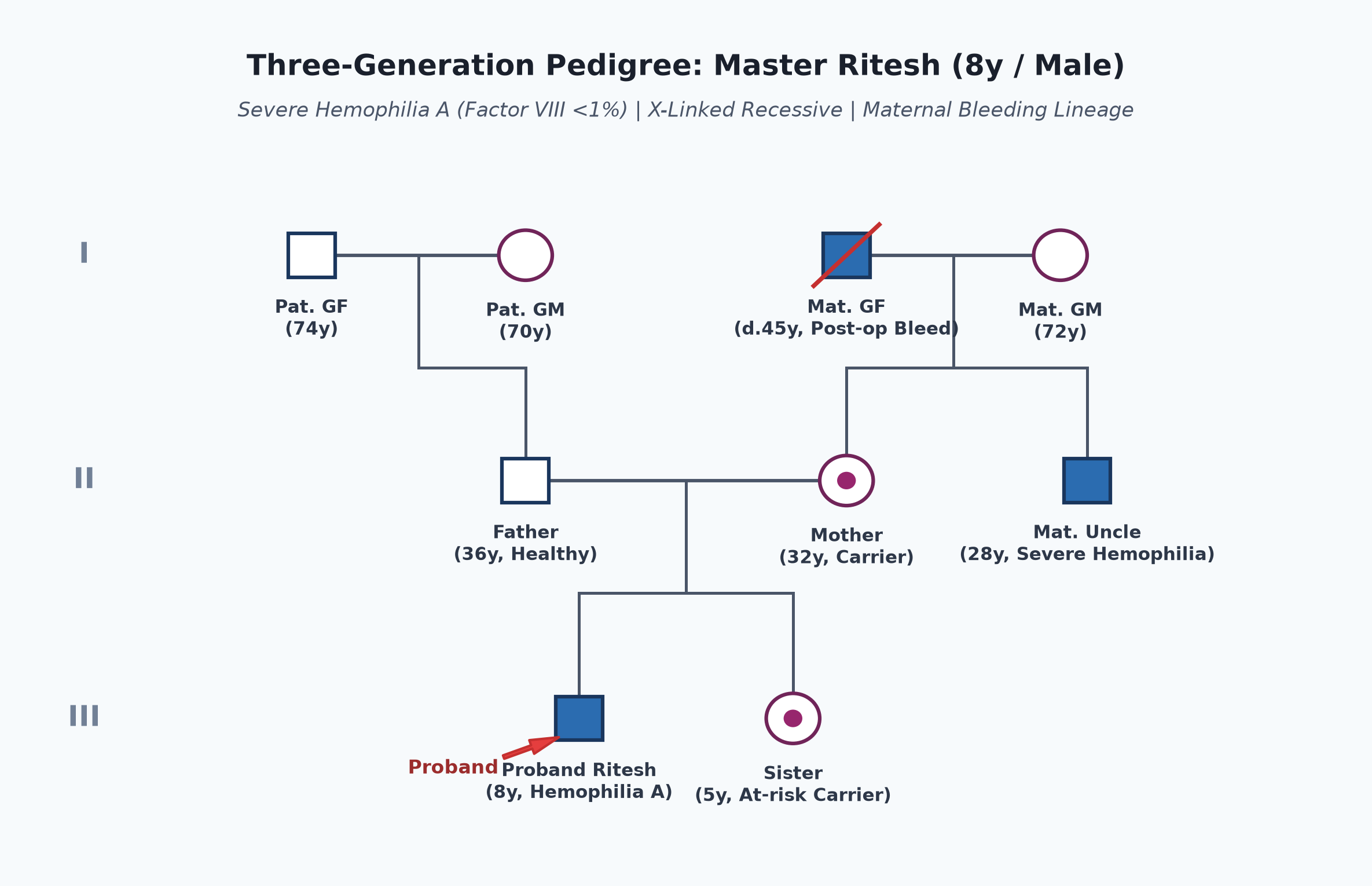
Immunization history
- Received all routine vaccines up to age as per the UIP.
- All vaccines were administered subcutaneously using a fine 25-gauge needle, followed by firm manual pressure for 5 minutes without rubbing (preventing deep intramuscular hematomas).
- Completely immunized against Hepatitis B (3 doses + booster) and Hepatitis A.
Dietary history
- Consumes a mixed northeastern Indian home diet consisting of rice, fish, eggs, and seasonal vegetables.
| Food Item | Quantity | Calories (kcal) | Protein (g) |
|---|---|---|---|
| Boiled Rice | 200 g | 260 | 5.2 |
| Fish Curry (Rohu) | 60 g fish | 80 | 11.5 |
| Dal | 1 bowl | 100 | 6.0 |
| Vegetable Stir Fry | 1 bowl | 90 | 1.8 |
| Boiled Egg (1) | 50 g | 75 | 6.2 |
| Total Observed Daily Intake | — | 605 kcal | 30.7 g |
24-Hour Recall Deficit Analysis
$$ \text{Ideal Body Weight (IBW for 8 years, 50th centile WHO)} = 25.3\text{ kg} $$| Nutrient | Expected Intake (ICMR-NIN 2024 for IBW 25.3 kg) | Observed Intake | Deficit | Percentage Deficit |
|---|---|---|---|---|
| Energy (kcal) | $25.3\text{ kg} \times 65\text{ kcal/kg} = 1645\text{ kcal}$ | 605 kcal | 1040 kcal | 63.2% Deficit |
| Protein (g) | $25.3\text{ kg} \times 1.0\text{ g/kg} = 25.3\text{ g}$ | 30.7 g | Nil (Adequate) | 0% Deficit |
The expected calories and proteins should be calculated from the ideal body weight, not from current weight.
Socioeconomic and KAP
- Belongs to Modified BG Prasad Socioeconomic Class III (Middle Class).
- Lives in Guwahati near a tertiary government hospital with an active Hemophilia Treatment Centre (HTC).
- Parents are well-educated regarding hemophilia; carry a Hemophilia Identity Card; seeking access to regular factor prophylaxis or Emicizumab under government schemes.
Summary of History
Master Ritesh, an 8-year-old boy, born of non-consanguineous parents with an X-linked maternal family history of bleeding disorders from Guwahati, presented with acute onset of painful tense swelling and functional disability of the right knee joint for 1 day following minor trauma, with a background history of recurrent hemarthroses involving the same right knee ("target joint"), delayed bleeding from lacerations, and prior factor VIII therapy, without superficial purpura, head trauma symptoms, or systemic bleeding.
I would like to think of an inherited coagulation factor deficiency, specifically Severe Hemophilia A (Factor VIII Deficiency), presenting with an Acute Right Knee Hemarthrosis superimposed on Chronic Hemophilic Arthropathy of the Right Knee (Target Joint), without evidence of life-threatening intracranial or internal hemorrhage.
General head to toe examination
- Child Behavioral State: Child is alert, cooperative, lying supine with right lower limb supported on a pillow, grimacing with movement due to right knee pain.
- Vitals:
- Pulse Rate: 96 beats/minute, regular, normal volume.
- Respiratory Rate: 20 breaths/minute, regular.
- Blood Pressure: $100/64\text{ mmHg}$ ($50^{\text{th}}$ centile for height and age).
- Temperature: $37.0^\circ\text{C}$ (afebrile).
- Anthropometry:
| Parameter | Observed | Expected (50th WHO) | Z-score / Centile | Inference |
|---|---|---|---|---|
| Weight | 24.8 kg | 25.3 kg | $50^{\text{th}}\text{ centile}$ | Normal nutritional status |
| Height | 127 cm | 128 cm | $50^{\text{th}}\text{ centile}$ | Normal stature |
- General Physical Findings:
- Crucial Negatives: NO pallor, NO icterus, NO petechiae, NO ecchymoses, NO mucosal bleeding, NO gingival bleeding, NO palpable purpura.
- Lymphadenopathy / Edema / Cyanosis / Clubbing: Completely absent.
- Oropharynx clear; no retropharyngeal bulge or airway compromise.
Systemic Examination
Musculoskeletal Examination: Right Knee Joint vs Normal Left Knee
- Inspection:
- Right lower limb held in an antalgic attitude of ~30 degrees flexion (position of maximum joint capsule capacity).
- Gross tense swelling of the entire right knee joint with complete obliteration of normal parapatellar depressions and suprapatellar pouch.
- Overlying skin is tense, smooth, and shiny; no open wounds, no cuts, no superficial ecchymoses.
- Muscle Wasting: Evident wasting of the right quadriceps muscle belly compared to the left thigh.
- Palpation:
- Marked local warmth over the right knee compared to the normal left knee.
- Intense tenderness elicited on gentle palpation over the joint line, patella, and suprapatellar pouch.
- Patellar Tap Test: Positive (patella floats and taps against the femoral condyle, confirming marked intra-articular effusion).
- Fluctuation Test: Cross-fluctuation across the joint space is positive.
- Periarticular synovial thickening palpable along the joint margins (chronic synovitis).
- Measurements (Comparative):
- Circumference at Mid-Patella: Right Knee = $31.5\text{ cm}$ | Left Knee = $26.8\text{ cm}$ ($+4.7\text{ cm}$ enlargement from acute hemarthrosis).
- Thigh Girth (10 cm above patella): Right Thigh = $31.0\text{ cm}$ | Left Thigh = $33.5\text{ cm}$ ($2.5\text{ cm}$ quadriceps atrophy due to chronic disuse).
- Range of Movement (ROM):
- Active and passive movements are severely restricted and excruciatingly painful; flexion beyond 30 degrees and extension to 0 degrees are resisted (fixed flexion deformity of ~5 degrees present).
- Other Joints: Left knee, bilateral ankles, elbows, and wrists show normal contour, no effusion, and full range of movement.
Central Nervous System (CNS)
- Conscious, oriented, clear speech.
- Cranial nerves I-XII clinically intact.
- Motor System (Upper Limbs & Left Lower Limb): Normal bulk, tone, and power (5/5); DTRs $2+$; plantars flexor bilaterally.
- Meningeal Signs: Absent. Fundoscopy: Normal discs, no retinal hemorrhages.
Abdomen & Other Systems
- Abdomen soft, non-tender; no hepatomegaly, no splenomegaly; renal angles non-tender.
- Psoas sign negative (no pain on hyperextension of hip, rules out iliopsoas hematoma).
- CVS & RS completely normal.
Summary
Master Ritesh, an 8-year-old boy, born of non-consanguineous parents with an X-linked maternal pedigree of bleeding disorders from Guwahati, presented with an acute onset of painful, hot, tense swelling and total immobility of the right knee joint for 1 day following a trivial stumble, on a background history of recurrent hemarthroses and target joint involvement. Examination demonstrates an acute right knee hemarthrosis (joint circumference $+4.7\text{ cm}$, positive patellar tap, local warmth, antalgic flexion), chronic quadriceps muscle atrophy ($2.5\text{ cm}$), and early arthropathy, without superficial petechiae, systemic bleeding, or neurological deficits.
The clinical findings are classic for Severe Hemophilia A (Congenital Factor VIII Deficiency), presenting with an Acute Right Knee Hemarthrosis involving a known Target Joint, with early Chronic Hemophilic Arthropathy.
Differential Diagnosis
| Disease | Points IN FAVOR | Points AGAINST |
|---|---|---|
| Severe Hemophilia A (Factor VIII Deficiency) | Male, X-linked maternal inheritance, deep hemarthrosis, target joint, prolonged aPTT with normal PT, Factor VIII <1% | Primary Diagnosis |
| Hemophilia B (Factor IX Deficiency / Christmas Disease) | Identical clinical presentation, deep hemarthroses, X-linked recessive | Indistinguishable clinically; differentiated strictly by specific factor assays (Factor IX deficient in Hemophilia B, normal in Hemophilia A) |
| Severe von Willebrand Disease (Type 3 / Severe Type 2) | Bleeding disorder, can present with hemarthrosis if FVIII is severely secondary depleted | Autosomal inheritance (affects both sexes equally), predominantly mucosal/cutaneous bleeding, prolonged Bleeding Time (BT) / abnormal PFA-100, markedly reduced vWF:Ag |
| Factor XI Deficiency (Hemophilia C) | Coagulation defect, prolonged aPTT | Autosomal recessive (equal sex ratio), spontaneous hemarthroses are exceptionally rare; bleeds only post-surgery/trauma |
| Septic Arthritis of the Knee | Acute painful knee swelling, local warmth, immobility | Associated with high spiking fever, toxic appearance, leukocytosis, elevated CRP/ESR; clear history of hemophilia and trauma points to hemarthrosis |
| Juvenile Idiopathic Arthritis (Oligoarticular JIA) | Monoarthritis of the knee joint in a child | Insidious onset, morning stiffness, lack of severe throbbing pain, normal coagulation parameters, no X-linked bleeding history |
Investigation Protocol & Diagnostic Workup
flowchart TD
    A["Male Child with Acute Hemarthrosis & Prolonged Bleeding"] --> B["Coagulation Screen: Platelet Count, PT/INR, aPTT & Bleeding Time"]
    B --> C{"Isolated Markedly Prolonged aPTT with Normal PT & Platelets?"}
    C -->|Yes| D["50:50 Mixing Study (Incubated Patient Plasma + Normal Plasma)"]
    C -->|No / Both Prolonged| E["Workup for Liver Disease, Vit K Deficiency, or DIC"]
    D --> F{"Complete Correction of aPTT to Normal?"}
    F -->|Corrects| G["Confirms Factor Deficiency: Measure Factor VIII:C & Factor IX:C"]
    F -->|Fails to Correct| H["Workup for Acquired Inhibitor / Lupus Anticoagulant (Bethesda Assay)"]
    G --> I{"Factor VIII:C < 1% with Normal Factor IX?"}
    I -->|Yes| J["Confirm Severe Hemophilia A: Screen for Inhibitors (< 0.4 BU) & MSK-USG"]
1. Coagulation Screening Battery
- Platelet Count: $285,000/\mu\text{L}$ (normal).
- Prothrombin Time (PT / INR): Normal ($12.2\text{ seconds}$, Control $12.0\text{ s}$, INR 1.0) — confirms extrinsic and common pathways are intact.
- Activated Partial Thromboplastin Time (aPTT): Markedly Prolonged at $78.0\text{ seconds}$ (Control $28-32\text{ seconds}$) — confirms an isolated intrinsic pathway coagulation defect.
- Bleeding Time (BT) / PFA-100 Closure Time: Normal (normal primary hemostasis; excludes von Willebrand disease and platelet dysfunction).
- Thrombin Time (TT): Normal ($15.0\text{ seconds}$).
2. 50:50 Plasma Mixing Study (Correction Test)
- Patient's plasma is mixed $1:1$ with normal pooled plasma and incubated at $37^\circ\text{C}$ for 2 hours.
- Result: aPTT completely corrects to normal ($30.5\text{ seconds}$).
- Inference: Confirms a true coagulation factor deficiency and excludes the presence of an immediate-acting or time-dependent circulating factor inhibitor.
3. Confirmatory Specific Coagulation Factor Assays
- Factor VIII Coagulant Activity (FVIII:C): $<1.0\%$ ($<0.01\text{ IU/mL}$) — Confirms Severe Hemophilia A.
- Severity Stratification:
- Severe: $<1\%$ of normal ($<0.01\text{ IU/mL}$) — spontaneous bleeds into joints and muscles.
- Moderate: $1-5\%$ of normal ($0.01-0.05\text{ IU/mL}$) — bleeds with minor trauma.
- Mild: $>5-40\%$ of normal ($0.05-0.40\text{ IU/mL}$) — bleeds only with major trauma or surgery.
- Severity Stratification:
- Factor IX Coagulant Activity (FIX:C): Normal at $92\%$ (excludes Hemophilia B).
- von Willebrand Factor Studies: vWF Antigen ($105\%$) and Ristocetin Cofactor Activity ($98\%$) are normal.
4. Inhibitor Screening
- Bethesda Assay (Nijmegen Modification): Negative ($<0.4\text{ Bethesda Units / BU}$) — confirms absence of neutralising anti-Factor VIII alloantibodies.
5. Imaging of the Affected Joint
- Point-of-Care Ultrasound (POCUS / MSK-USG Right Knee):
- Demonstrates marked anechoic to complex hypoechoic suprapatellar joint effusion with thickened, hypervascular synovial folds (HEAD-US score $4/8$).
- Plain Radiograph of Right Knee (AP and Lateral):
- Demonstrates soft tissue distension, periarticular osteopenia, widening of the intercondylar notch, and subtle subchondral irregularity (Arnold-Hilgartner Stage II Hemophilic Arthropathy).
Management Plan
1. Acute Hemarthrosis: Immediate Factor VIII Replacement Therapy
- Treatment Principle: Treat immediately upon suspicion; NEVER delay factor infusion for radiological imaging or laboratory confirmation.
- Factor VIII Dose Calculation Formula:
- For an Acute Hemarthrosis, the target desired factor rise is $40\% - 60\%$.
- Calculation for Master Ritesh (Weight 25.0 kg):
- Prescription: Administer $650\text{ Units}$ (or $25\text{ U/kg}$) of Recombinant or Plasma-Derived Factor VIII Concentrate as an immediate intravenous push over 5 minutes.
- Maintenance Dosing: Re-dose at $20-25\text{ U/kg}$ every 12 hours for 24–48 hours until acute pain, warmth, and joint swelling significantly regress.
2. Adjunctive Joint Management: The PRICE Protocol
- P — Protection: Splint the right knee in comfortable 20-30 degree antalgic flexion using a padded posterior plaster slab or removable knee brace.
- R — Rest: Strict bed rest; avoid all weight bearing on the affected limb.
- I — Ice: Apply cold packs wrapped in a towel for 15–20 minutes every 4 to 6 hours (causes vasoconstriction and numbs pain).
- C — Compression: Gentle elastic crepe bandage wrapping (avoid tight circumferential constriction).
- E — Elevation: Elevate the right lower limb on pillows above heart level to enhance venous and lymphatic drainage.
- Analgesia: Prescribe Oral Paracetamol ($15\text{ mg/kg/dose}$ q6h) or Tramadol for severe pain. STRICTLY PROHIBIT Aspirin and NSAIDs (Ibuprofen/Diclofenac) due to irreversible platelet inhibition.
3. Rehabilitation & Physiotherapy
- As soon as acute pain and swelling subside under factor coverage (usually day 2–3), initiate supervised isometric quadriceps strengthening exercises and active-assisted range of motion.
- Clinical Rationale: Prompt rehabilitation prevents arthrofibrosis, capsular contracture, and worsening quadriceps atrophy.
4. Long-Term Regular Prophylaxis (Standard of Care - WFH 2020 Guidelines)
- Transition from episodic "on-demand" treatment to Regular Primary/Secondary Prophylaxis:
- Standard Factor VIII Prophylaxis: Intravenous Recombinant Factor VIII at $25-40\text{ U/kg}$ administered 3 times weekly (or every other day) to maintain trough factor VIII levels consistently $>1-3\%$.
- Novel Non-Factor Prophylaxis (Emicizumab - Hemlibra):
- Humanized bispecific monoclonal antibody that bridges activated Factor IX and Factor X, restoring the function of missing Factor VIII.
- Dosage: $3.0\text{ mg/kg}$ subcutaneously once weekly for 4 weeks (loading dose), followed by maintenance therapy of $1.5\text{ mg/kg}$ subcutaneously once weekly (or $3.0\text{ mg/kg}$ every 2 weeks).
- Advantages: Subcutaneous administration (eliminates venous access difficulties), constant hemostatic protection, and equal efficacy in patients with or without Factor VIII inhibitors.
5. Comprehensive Hemophilia Care & Family Guidance
- Provide a durable Hemophilia Medical Alert Card / Bracelet indicating diagnosis, factor deficiency, and emergency contact numbers.
- Avoid Intramuscular Injections: All future immunizations and medications must be administered subcutaneously or orally.
- Dental Hygiene: Meticulous dental care to prevent caries and avoid dental extractions; co-prescribe oral Tranexamic acid ($15-20\text{ mg/kg/dose}$ tid for 5 days) for any minor mucosal or dental oozing (antifibrinolytic).
- Family Screening: Provide genetic counseling; perform carrier testing for the mother and 5-year-old younger sister.