
Master Tanmay, a 3-year-old male child, only child born of a non-consanguineous marriage from Patna, Bihar, presented with complaints of an asymptomatic, rapidly enlarging swelling in the right side of the abdomen noticed by his mother during bathing 3 weeks ago, low-grade intermittent fever for 10 days, and microscopic hematuria noted on routine urine testing, with the child appearing otherwise energetic, robust, playful, and without any bone pain, periorbital ecchymosis, jaundice, or bowel obstruction.
- Asymptomatic abdominal mass accidentally discovered by parents while bathing or dressing ('the well child with a large belly')
- Abdominal pain or dull flank ache (present in 30-40% due to subcapsular hemorrhage or stretching)
- Gross or microscopic hematuria (present in 20-30% due to invasion into renal pelvicalyceal system)
- Systemic hypertension (present in 25-30% due to renal ischemia and hyperreninemia)
- Low-grade fever (present in 20% due to tumor necrosis)
- Striking ABSENCE of cachexia, severe bone pain, or systemic prostration seen in neuroblastoma
HOPI
The child was in excellent health until 3 weeks ago when his mother, while bathing him, noticed a firm, painless bulge in the right flank and upper abdomen.
An abdominal lump in a preschooler is one of the most classic surgical-pediatric exam cases. The primary clinical dilemma is distinguishing Wilms Tumor (Intrarenal Nephroblastoma) from Neuroblastoma (Retroperitoneal Adrenal Medullary Neoplasm). Inquire meticulously regarding the child's general well-being: children with Wilms tumor typically look remarkably robust, thriving, and happy ('the healthy baby with an abdominal mass'), whereas children with neuroblastoma are characteristically pale, miserable, irritable, wasting, and suffering from metastatic bone pain. Specifically ask about periorbital bruising ('raccoon eyes'), opsoclonus-myoclonus (dancing eyes/feet), and flushing or diarrhea (secretory vasoactive intestinal peptide [VIP]).
- Asymptomatic Abdominal Mass:
- Accidentally palpated 3 weeks ago; has noticeably expanded over the past month.
- Not associated with crying on touch, vomiting, or abdominal distension.
- Microscopic Hematuria:
- Mother noticed urine was occasionally smoky or dark-amber in color 10 days ago.
- No burning sensation, dysuria, or graveluria.
- Low-Grade Intermittent Fever:
- Documented over the past 10 days, spiking to $100.2^\circ\text{F}$ ($37.9^\circ\text{C}$); non-toxic appearance.
- Systemic Negative Inquiries:
- No periorbital swelling, dark circles, or proptosis (rules out retro-orbital metastatic neuroblastoma).
- No bone pain, limp, or reluctance to run.
- No chronic watery diarrhea or profuse sweating (rules out VIP-secreting neuroblastoma).
- No hemihypertrophy, tongue enlargement, or umbilical hernia noted since birth (rules out Beckwith-Wiedemann syndrome).
- No aniridia or ambiguous genitalia (rules out WAGR syndrome).
Past history
- No history of previous urinary tract infections, renal stones, or abdominal trauma.
- No history of prior hospitalizations.
Antenatal, natal and postnatal history
- Mother was a 26-year-old primigravida; uneventful pregnancy; anomaly ultrasound at 20 weeks reported normal fetal kidneys and amniotic fluid index.
- Full-term normal delivery at hospital; birth weight 3.2 kg; cried immediately.
- Neonatal period uneventful; no hypoglycemia.
Development history
- Completely normal gross motor, fine motor, language, and social milestones; fully age-appropriate.
Family history
- Non-consanguineous marriage.
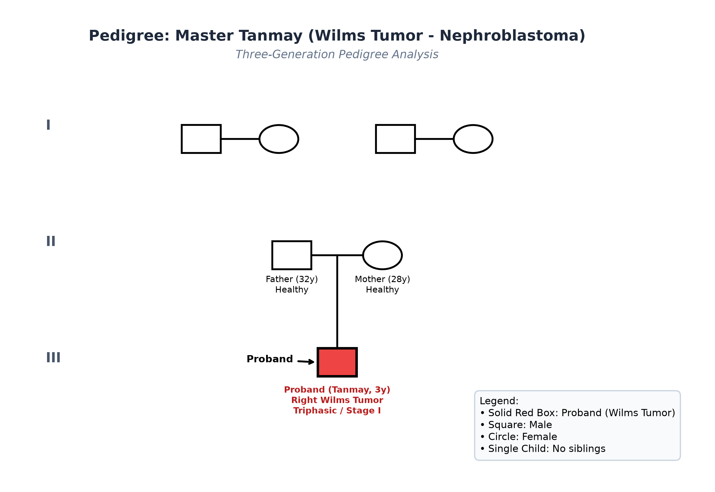
- Father 32 years, private executive, healthy; Mother 28 years, homemaker, healthy.
- No family history of childhood renal tumors, sarcomas, or early cancers.
Immunization history
- Fully immunized for age per the National Immunization Schedule.
Dietary history
- Consumes an age-appropriate balanced diet; appetite is preserved.
| Food Item | Quantity | Calories (kcal) | Protein (g) |
|---|---|---|---|
| Cow's Milk (toned) | 300 mL | 180 | 9.6 |
| Roti (whole wheat, 1.5) | 45 g flour | 153 | 5.2 |
| Boiled Rice | 100 g cooked | 130 | 2.6 |
| Dal (Toor dal, 1 katori) | 30 g raw | 102 | 6.6 |
| Cooked Mixed Sabzi | 1 bowl | 110 | 2.2 |
| Banana | 1 medium (80 g) | 72 | 1.0 |
| Ghee (in dal/roti) | 5 g | 45 | 0.0 |
| Snacks / Biscuit | 30 g | 120 | 2.1 |
| Total Observed Daily Intake | — | 912 kcal | 29.3 g |
24-Hour Recall Deficit Analysis
$$ \text{Ideal Body Weight (IBW for 3 years, 50th centile WHO)} = 14.3\text{ kg} $$| Nutrient | Expected Intake (ICMR-NIN 2024 for IBW 14.3 kg) | Observed Intake | Deficit | Percentage Deficit |
|---|---|---|---|---|
| Energy (kcal) | $14.3\text{ kg} \times 78\text{ kcal/kg} = 1115\text{ kcal}$ | 912 kcal | 203 kcal | 18.2% Deficit (Mild) |
| Protein (g) | $14.3\text{ kg} \times 1.0\text{ g/kg} = 14.3\text{ g}$ | 29.3 g | Nil (Adequate) | 0% Deficit |
The expected calories and proteins should be calculated from the ideal body weight, not from current weight.
Socioeconomic and KAP
- Modified BG Prasad Socioeconomic Class II (Upper Middle Class).
- Lives in a well-ventilated urban pucca apartment.
Summary of History
Master Tanmay, a 3-year-old thriving male child, presents with a 3-week history of an accidentally discovered, rapidly enlarging, painless right flank abdominal mass accompanied by low-grade fever, microscopic hematuria, and preserved appetite, without bone pain, diarrhea, or dysmorphic syndrome features.
I would like to consider a provisional diagnosis of Pediatric Solid Abdominal Tumor (most consistent with Wilms Tumor / Nephroblastoma of the Right Kidney), clinically Stage I/II, in a well-nourished child.
General head to toe examination
- General Appearance: Alert, playful, robust, well-nourished child, sitting comfortably in bed.
- Vitals:
- Heart Rate: 104 beats/minute, regular.
- Respiratory Rate: 22 breaths/minute, regular.
- Blood Pressure: $116/76\text{ mmHg}$ ($>95^{\text{th}}\text{ percentile} + 12\text{ mmHg}$ for 3-year-old male at $50^{\text{th}}$ height centile $\to$ Stage 2 Systemic Hypertension).
- Temperature: $37.2^\circ\text{C}$ ($99.0^\circ\text{F}$) axillary.
- Capillary Refill Time: $<2$ seconds.
- Anthropometry:
| Parameter | Observed | Expected (50th WHO) | Z-score / Centile | Inference |
|---|---|---|---|---|
| Weight | 14.8 kg | 14.3 kg | $+0.2\text{ SD}$ | Normal Weight (Robust) |
| Pre-morbid Weight | 14.5 kg | 14.3 kg | $50^{\text{th}}\text{ centile}$ | Stable baseline weight |
| Height | 96.0 cm | 96.1 cm | $50^{\text{th}}\text{ centile}$ | Normal Stature |
| Weight-for-Height | 14.8 kg for 96 cm | 14.3 kg | $+0.4\text{ SD}$ | Well-nourished |
| Head Circumference | 49.2 cm | 49.5 cm | Normal | Age-appropriate |
| BMI | $16.0\text{ kg/m}^2$ | $15.5\text{ kg/m}^2$ | $+0.4\text{ SD}$ | Well-nourished |
- Dysmorphic Screening:
- Eyes: Torch and slit-lamp examination: normal irides bilaterally (no aniridia; excludes WAGR syndrome).
- Face & Oral: Normal tongue size (no macroglossia), no anterior ear lobe pits or helical creases (excludes Beckwith-Wiedemann syndrome).
- Limbs: Symmetrical length and girth of upper and lower limbs (no hemihypertrophy).
- Orbits: Normal globes, no periorbital ecchymosis, no proptosis, no Horner syndrome.
- Lymphadenopathy: Complete absence of palpable cervical, axillary, or supraclavicular lymphadenopathy.
- Pallor: Mild pallor present.
Detailed Abdominal Examination
NEVER repeatedly or vigorously palpate a suspected Wilms tumor! Palpation must be extremely gentle. Heavy pressure can rupture the tense pseudo-capsule, spilling tumor cells into the peritoneal cavity and immediately transforming a resectable Stage I/II tumor into an advanced Stage III tumor requiring whole-abdominal radiation!
Inspection
- Asymmetrical fullness visible in the right lumbar and hypochondriac regions.
- Overlying skin is normal; no visible venous collateral engorgement or peristalsis.
- Umbilicus is central, normal, no umbilical hernia.
Palpation
- Location: Mass occupies the right lumbar, right hypochondriac, and extends into the right iliac fossa.
- Dimensions: Measures approximately $10.0\text{ cm} \times 8.0\text{ cm}$.
- Surface & Consistency: Firm, smooth, uniform, globular surface with rounded margins; completely non-tender.
- Mobility: Moves slightly with deep inspiration.
- Midline Relationship (CRITICAL PHYSICAL SIGN): Medial border is well-defined, situated approximately 2.0 cm to the right of the midline $\to$ THE MASS DOES NOT CROSS THE MIDLINE!
- Bimanual Palpation & Ballottement:
- Posteriorly fills the renal angle; Bimanually palpable and distinctly Ballotable $\to$ Confirms Intrarenal Retroperitoneal Origin.
- Normal left kidney; no other palpable mass or hepatosplenomegaly.
Percussion & Auscultation
- Resonant note of colonic gas heard anterior to the medial border of the mass (Band of colonic resonance positive); dull over the flank.
- Bowel sounds normal; no renal bruit audible over the mass.
Summary
Master Tanmay, a 3-year-old thriving male child, presents with an accidentally detected, smooth, firm, non-tender, ballotable right renal mass measuring $10 \times 8\text{ cm}$ that does not cross the midline, accompanied by Stage 2 systemic hypertension and microscopic hematuria, without syndromic features (aniridia, hemihypertrophy), metastases, or cachexia.
Final Clinical Diagnosis: Pediatric Solid Abdominal Tumor: Wilms Tumor (Nephroblastoma) of the Right Kidney, clinically Stage I/II, presenting with renin-mediated secondary hypertension.
Differential Diagnosis: Wilms vs Neuroblastoma
| Diagnostic Feature | Wilms Tumor (Nephroblastoma) | Abdominal Neuroblastoma |
|---|---|---|
| Age of Onset | Peak 3 to 4 years (Tanmay is 3 years) | Younger: median 18 months; usually $<2$ years |
| General Health | Robust, thriving, well-nourished | Sick, toxic, cachectic, bone pain, irritable |
| Origin | Intrarenal (Renal blastema) | Extrarenal (Adrenal medulla / sympathetic chain) |
| Consistency & Surface | Firm, smooth, uniform, globular | Stony-hard, nodular, irregular, bosselated |
| Midline Crossing | DOES NOT CROSS MIDLINE (confined to renal fossa) | CROSSES MIDLINE (encases aorta/IVC/mesenteric vessels) |
| Ballottement | Ballotable (pushes colon medially) | Non-ballotable, fixed to retroperitoneum |
| Hypertension | Present in $25-30\%$ (Renin hypersecretion) | Present in $25\%$ (Catecholamine hypersecretion) |
| Metastatic Sites | Lungs (#1), Liver | Bone (#1), Bone marrow, Orbits (Raccoon eyes), Liver |
| Urinary Markers | Normal VMA/HVA | Elevated Urinary VMA and HVA ($>90\%$) |
| Radiology (CT/USG) | 'Claw Sign' / parenchyma cap; rare calcifications | Coarse calcifications ($>85\%$), encases vessels without invasion |
Investigation Protocol & Diagnostic Workup
flowchart TD
    A["Preschool Child with Smooth, Non-Tender, Ballotable Flank Mass"] --> B["Abdominal Ultrasound with Doppler: Origin & Renal Vein/IVC Patency"]
    B --> C["Contrast-Enhanced CT (CECT) of Abdomen & Pelvis"]
    C --> D{"Confirms Intrarenal Origin with 'Claw Sign' & Intact Capsule?"}
    D -->|Yes| E["Chest CT (High-Resolution): Rule Out Pulmonary Metastases (Stage IV)"]
    E --> F["Urine Analysis (Hematuria) & 24h Urine VMA/HVA (Exclude Neuroblastoma)"]
    F --> G["Determine Treatment Philosophy: COG/NWTS vs SIOP Protocol"]
    G --> H["COG Approach: Upfront Radical Nephrectomy + Adjuvant Chemotherapy"]
    G --> I["SIOP Approach: Neoadjuvant Vincristine + Dactinomycin x 4 wk -> Surgery"]
1. Imaging Modalities
- Abdominal Ultrasound with Doppler:
- Large well-defined, heterogeneous mass measuring $10.2 \times 8.1\text{ cm}$ arising from the mid and lower poles of the right kidney.
- Classic 'Claw Sign' (compressed normal renal parenchyma capping the tumor).
- Doppler shows patent right renal vein and inferior vena cava (IVC) with no tumor thrombus.
- Left kidney is completely normal (excludes bilateral Stage V disease).
- Contrast-Enhanced CT (CECT) of Abdomen & Pelvis:
- Confirms a solid, circumscribed, heterogeneous intrarenal mass with areas of internal necrosis.
- Renal capsule is intact without perinephric fat infiltration.
- Abdominal lymph nodes $<1.0\text{ cm}$ (no lymphadenopathy).
- High-Resolution CT (HRCT) of Chest:
- Lung fields clear; no pulmonary cannon-ball metastases (excludes Stage IV disease).
2. Laboratory & Biomarker Diagnostics
- Serum Creatinine: $0.5\text{ mg/dL}$, Blood Urea: $18\text{ mg/dL}$ (Normal renal function).
- Complete Blood Count: Hb $10.8\text{ g/dL}$, TLC $8,400/\mu\text{L}$, Platelets $320,000/\mu\text{L}$.
- Urine Analysis: Microscopic hematuria ($15-20\text{ RBCs/HPF}$); protein negative.
- Urinary Catecholamine Metabolites (VMA and HVA): Normal (firmly rules out Neuroblastoma).
Multimodal Management Plan
1. Antihypertensive Therapy
- Start oral Calcium Channel Blocker: Amlodipine ($0.1\text{ to } 0.2\text{ mg/kg/day}$ orally once daily). (Avoid ACE inhibitors upfront if renal artery stenosis or severe contralateral hypoperfusion is unverified).
2. Surgical Resection: Upfront Radical Nephrectomy (COG / NWTS Protocol)
- Approach: Transabdominal transperitoneal transverse laparotomy (allows full visualization of contralateral kidney and great vessels).
- Steps:
- Early ligation of the right renal vein and artery to prevent intraoperative hematogenous tumor embolization.
- Complete en-bloc resection of the right kidney, tumor, perinephric fat, and Gerota's fascia.
- Mandatory Regional Lymph Node Sampling: Biopsy of para-aortic and paracaval lymph nodes (even if clinically normal) for definitive staging.
- Inspection and palpation of the contralateral left kidney.
- Strict Rule: Avoid capsule rupture; if spillage occurs, staging escalates immediately to Stage III!
3. Pathological Staging & Risk Stratification (NWTS System)
- Stage I: Tumor limited to kidney, capsule intact, completely resected, negative margins and lymph nodes.
- Stage II: Extends beyond kidney (into perirenal fat or renal sinus vessels) but completely resected with clear margins.
- Stage III: Residual non-hematogenous tumor in abdomen (rupture, positive margins, or positive lymph nodes).
- Stage IV: Distant hematogenous metastases (Lungs, liver).
- Stage V: Bilateral renal involvement.
- Histology: Favorable Histology (FH, triphasic: blastemal, epithelial, stromal without anaplasia).
- Molecular Markers: Evaluate for $1p$ and $16q$ Loss of Heterozygosity (LOH).
4. Adjuvant Chemotherapy
- For Stage I / II Favorable Histology:
- Two-Drug Chemotherapy Regimen: Vincristine ($1.5\text{ mg/m}^2$ IV) + Dactinomycin / Actinomycin D ($0.045\text{ mg/kg}$ IV) administered for 18 weeks.
- Overall survival exceeds $95\%$ with this regimen, without requiring radiotherapy!
- Radiotherapy: Omitted in Stage I and II Favorable Histology; indicated only for Stage III (flank RT) and Stage IV (whole lung RT).