
Master Sourav, an 8 year old boy, 1st order child born of a non-consanguineous marriage from Kolkata, West Bengal presented with complaints of continuous, low-to-moderate grade fever with evening chills since 4 weeks, progressive pallor and weight loss since 3 weeks, left-sided upper abdominal pain since 4 days, and painful red spots on the fingertips since 2 days.
- Prolonged, unexplained fever in a child with known congenital or rheumatic heart disease
- Malaise, anorexia, weight loss, and progressive pallor (anemia of chronic disease)
- Peripheral immunological or embolic stigmata (splinter hemorrhages, Osler nodes, Janeway lesions, Roth spots)
- Acute worsening of an existing murmur, new-onset murmur, or development of heart failure
HOPI
The history is dated back to 4 weeks ago when the child developed insidious onset of low-to-moderate grade fever.
In every child presenting with prolonged pyrexia and a heart murmur, maintain a high index of suspicion for IE. Probe specifically for: 1) Underlying cardiac substrate (CHD, RHD, prosthetic material); 2) Portals of bacteremic entry (dental extraction, skin sepsis, central venous lines); 3) Embolic phenomena (stroke, hematuria, splenic infarction); 4) Immunological manifestations.
- Fever & Constitutional Symptoms:
- Low-to-moderate grade ($38.2^\circ ext{C}$ to $38.8^\circ ext{C}$), documented daily for 4 weeks.
- Associated with chills and evening sweating.
- Temporarily subsided with oral paracetamol and multiple outpatient courses of oral amoxicillin-clavulanate and cefixime, but recurred promptly upon stopping antibiotics Partially treated endocarditis with suppressed bacteremia and delayed diagnosis.
- Associated with marked anorexia, profound lethargy, and loss of 2.5 kg weight over 4 weeks.
- Portal of Entry (Dental Procedure):
- History of a dental extraction for severe dental caries performed 6 weeks ago at a local clinic without prior antibiotic prophylaxis.
- Peripheral Embolic & Immunological Phenomena:
- Painful, tender, red, pea-sized raised nodules appeared over the pulp of the left index and middle fingers 2 days ago (Osler nodes) Immunological immune complex deposition in dermal microvessels.
- Mother noticed tiny dark-red linear streaks underneath two fingernails 5 days ago (Splinter hemorrhages).
- No painless hemorrhagic macules on the palms or soles (Janeway lesions).
- Abdominal Pain (Splenic Infarction):
- Developed sudden, sharp, localized left upper quadrant abdominal pain 4 days ago, worsening on deep inspiration and radiating to the left shoulder Acute splenic infarction from systemic embolization of left-sided vegetative fragments.
- Worsening Cardiac Status:
- Mother reports that over the past week, the child has developed shortness of breath on walking and orthopnea, which were not present previously Acute valvar incompetence or rupture of chordae causing new-onset heart failure.
- Negative History:
- No history of sudden loss of consciousness, focal seizures, speech difficulty, or weakness of one half of the body Rules out cerebral mycotic aneurysm rupture or embolic stroke.
- No history of gross hematuria (cola-colored urine) or facial puffiness Rules out immune-complex mediated glomerulonephritis.
- No history of visual disturbance or sudden blindness Rules out retinal artery embolization or Roth spots affecting central vision.
- No history of central cyanosis or cyanotic spells.
Past History
- Known case of congenital heart disease (repaired Perimembranous VSD with residual small leak and mild aortic regurgitation / Bicuspid Aortic Valve) diagnosed at 2 years of age; lost to regular cardiology follow-up for 3 years.
- No history of previous episodes of infective endocarditis.
Family history
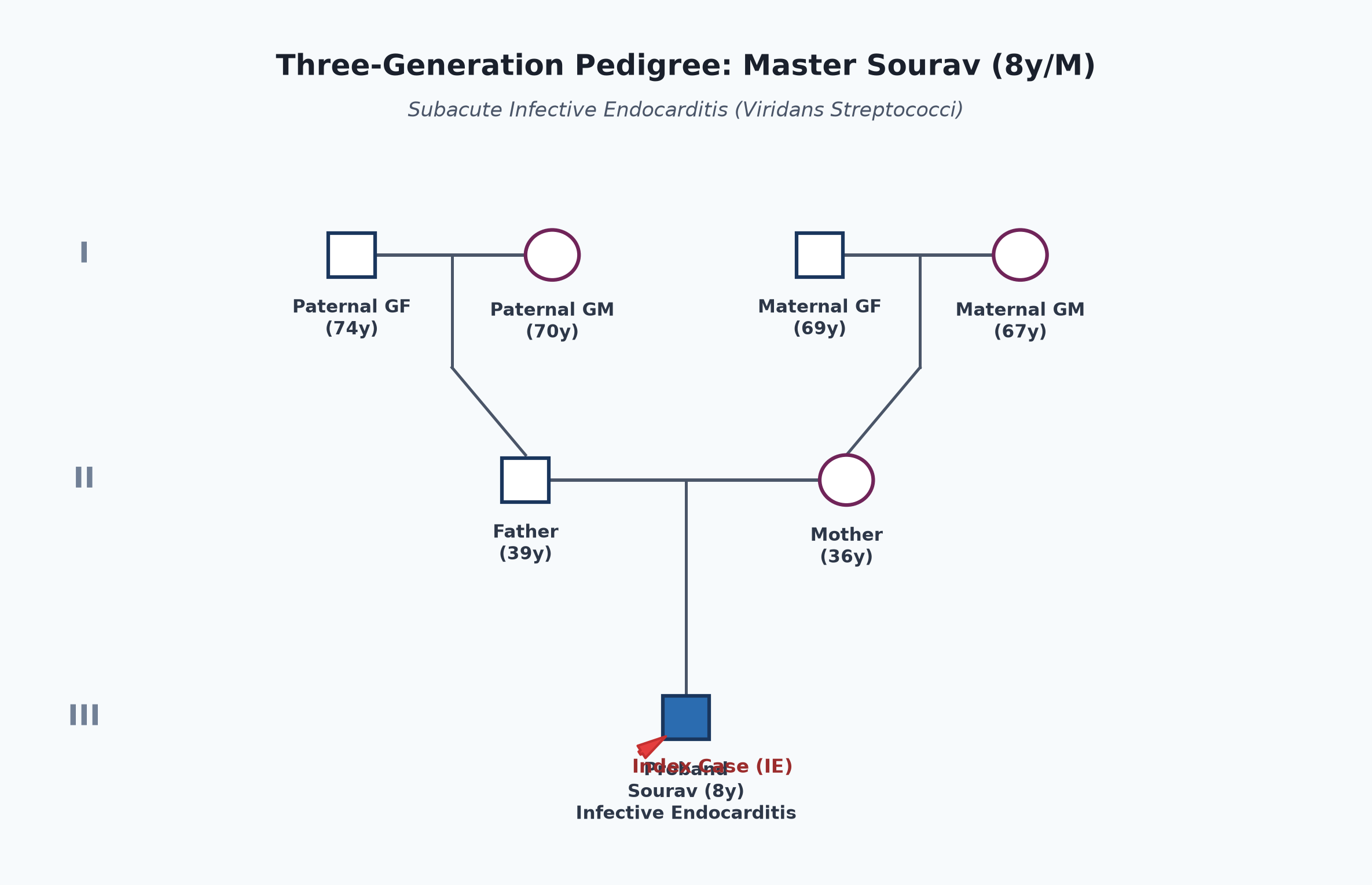
- Firstborn child of non-consanguineous parents.
- Father 39 years, teacher; mother 36 years, bank clerk. Both healthy.
- No family history of early heart disease or endocarditis.
Immunization history
- Fully immunized according to UIP up to 5 years.
- Received pneumococcal vaccine (PCV13).
Dietary history
- Normal mixed diet prior to illness; significant reduction in intake over past 4 weeks due to severe anorexia.
| Food Item | Quantity | Calories (kcal) | Protein (g) |
|---|---|---|---|
| Milk with cereal | 200 mL | 180 | 6.5 |
| Rice with boiled egg | 1/2 bowl rice + 1 egg | 220 | 8.2 |
| Khichdi / Soup | 1 small bowl | 150 | 4.0 |
| Fruit juice (fresh) | 100 mL | 55 | 0.5 |
| Total Observed Intake | — | 605 kcal | 19.2 g |
24-Hour Recall Deficit Analysis
$$ ext{Ideal Body Weight (IBW for 8-year-old boy, 50th centile WHO)} = 25.5 ext{ kg} $$| Nutrient | Expected Intake (ICMR-NIN 2024 for 7-9 years) | Observed Intake | Deficit | Percentage Deficit |
|---|---|---|---|---|
| Energy (kcal) | $1700 ext{ kcal/day}$ | 605 kcal | 1095 kcal | 64.4% Deficit |
| Protein (g) | $23.0 ext{ g/day}$ | 19.2 g | 3.8 g | 16.5% Deficit |
The expected calories and proteins should be calculated from the ideal body weight, not from current weight.
Socioeconomic and KAP
- Belongs to Modified BG Prasad Socioeconomic Class II (Upper Middle Class).
- Urban apartment with good sanitation.
- Parents were unaware that a child with a congenital heart defect requires prophylactic antibiotics and strict oral hygiene prior to invasive dental extractions.
Summary of History
Master Sourav, an 8-year-old male child with a known background of congenital heart disease (residual VSD / bicuspid aortic valve), presented with 4 weeks of continuous fever with chills following an unprophylaxed dental extraction, progressive pallor, weight loss, left hypochondriac pain, painful nodules on fingertips (Osler nodes), and recent-onset exertional dyspnea, without focal neurological deficits.
I would like to think of Subacute Infective Endocarditis (IE) involving the aortic valve and residual ventricular septal defect, complicated by splenic infarction and mild congestive cardiac failure, with prominent peripheral immunological and embolic stigmata.
General head to toe examination
- Child Behavioral State: Calm, conscious, cooperative, appears chronically sick and pale (Prechtl State 3).
- Vitals:
- Heart Rate: 124 beats/minute, regular, collapsing / water-hammer volume with rapid upstroke and rapid descent (due to acute/subacute aortic regurgitation); all peripheral pulses palpable; locomotor brachialis positive.
- Respiratory Rate: 28 breaths/minute, regular, mild intercostal retractions.
- Blood Pressure: Right arm supine: $118/42 ext{ mmHg}$ (wide pulse pressure of $76 ext{ mmHg}$; Hill's sign positive with popliteal cuff pressure $35 ext{ mmHg}$ higher than brachial).
- Temperature: $38.4^\circ ext{C}$ (febrile).
- Oxygen Saturation ($SpO_2$): 96% on room air.
- Anthropometry:
| Parameter | Observed | Expected (50th WHO) | Z-score / Centile | Inference |
|---|---|---|---|---|
| Weight | 20.8 kg | 25.5 kg | $-2.1 ext{ SD}$ | Moderate Underweight |
| Height | 125 cm | 128.0 cm | Normal | Normal Stature |
| BMI | $13.3 ext{ kg/m}^2$ | $15.5 ext{ kg/m}^2$ | $< 5^{ ext{th}} ext{ centile}$ | Acute Wasting |
- Head to Toe Findings:
- Pallor: Marked conjunctival, palmar, and nail bed pallor.
- Clubbing: Grade II clubbing present on fingers (Lovibond angle obliterated, positive fluctuation).
- Cutaneous Stigmata of Endocarditis:
- Osler Nodes: Tender, erythematous, raised pea-sized nodules palpable on the pulps of the left index and middle fingers.
- Splinter Hemorrhages: Linear, dark-red subungual streaks located in the distal third of the left thumbnail and right index fingernail.
- Petechiae: Conjunctival petechiae present in the lower palpebral conjunctiva bilaterally.
- Janeway lesions absent.
- Ophthalmoscopy (Fundus):
- Roth Spots: Oval retinal hemorrhages with pale, white fibrinous centers observed in the left superior retinal quadrant on direct fundoscopy.
- Oral Cavity: Evidence of multiple restored teeth and poor periodontal hygiene.
Systemic Examination
Cardiovascular System
- Inspection:
- Apical impulse visible in the left $5^{ ext{th}}$ intercostal space, $1.5 ext{ cm}$ lateral to the midclavicular line.
- Precordial hyperactivity present; visible carotid pulsations (Corrigan sign).
- Palpation:
- Apical Impulse: Palpable in the left $5^{ ext{th}}$ intercostal space, $1.5 ext{ cm}$ lateral to the midclavicular line; hyperdynamic and displaced downwards and outwards.
- Thrill: High-pitched early diastolic thrill palpable along the left $3^{ ext{rd}}$ and $4^{ ext{th}}$ intercostal spaces (Erb's point) with patient sitting forward in full expiration; systolic thrill at left lower sternal border.
- Grade 1/3 parasternal heave palpable.
- Auscultation:
- First Heart Sound ($S_1$): Normal intensity.
- Second Heart Sound ($S_2$): Soft aortic component ($A_2$); $P_2$ normal.
- Murmur 1 (New / Worsening Aortic Regurgitation):
- Grade 3/6, high-pitched, decrescendo early diastolic murmur (EDM) heard best at the $3^{ ext{rd}}$ left intercostal space (Erb's point) with the diaphragm, radiating along the left sternal border to the apex, elicited with the patient sitting forward in held expiration.
- Murmur 2 (Residual VSD Jet):
- Grade 3/6 harsh pansystolic murmur at the left $4^{ ext{th}}$ intercostal space with radiation.
- Austin Flint Murmur: Soft, low-pitched mid-diastolic murmur audible at the cardiac apex (regurgitant aortic jet impinging on the anterior mitral leaflet, creating functional mitral stenosis).
- Peripheral Vascular Signs of AR:
- Water-hammer (Corrigan) pulse, pistol-shot sounds over femoral arteries (Traube sign), and systolic/diastolic murmurs on light femoral compression (Duroziez sign).
other systems
- Abdomen:
- Spleen: Palpable 3.5 cm below the left costal margin in the anterior axillary line; firm, moderately tender, with a soft splenic friction rub audible on auscultation (confirms acute splenic infarction).
- Liver: Palpable 2 cm below right costal margin, soft, mildly tender.
- No ascites.
- Respiratory System: Bilateral vesicular breath sounds; scattered basal crackles.
- Central Nervous System: Normal sensorium, cranial nerves intact, no focal neurological deficits, no signs of meningeal irritation.
Summary
Master Sourav, an 8-year-old male child with a known background of congenital heart disease (repaired VSD with residual leak / bicuspid aortic valve), presented with 4 weeks of prolonged pyrexia with chills following an unprophylaxed dental extraction, marked pallor, Osler nodes, splinter hemorrhages, Roth spots, and left upper quadrant pain due to splenic infarction.
On examination, he has high-grade fever, wide pulse pressure ($76 ext{ mmHg}$) with peripheral signs of severe aortic regurgitation, a hyperdynamic displaced apex, a new Grade 3/6 early diastolic murmur at Erb's point, a Grade 3/6 pansystolic murmur at LLSB, Roth spots on fundoscopy, tender splenomegaly with a splenic rub, and mild hepatomegaly.
Probable Clinical Diagnosis: Definite Subacute Infective Endocarditis (IE) by Modified Duke Criteria (2 Major: oscillating intracardiac vegetation on echocardiogram + typical blood culture; 4 Minor: predisposing cardiac lesion, fever $>38^\circ ext{C}$, vascular/embolic phenomena [splenic infarct, splinter hemorrhages], immunological phenomena [Osler nodes, Roth spots]), with Acute-on-Chronic Severe Aortic Regurgitation and residual VSD, complicated by Splenic Infarction and mild heart failure, most likely caused by Viridans group Streptococci (Streptococcus sanguinis / oralis).
Differential Diagnosis
| Disease | Points IN FAVOR | Points AGAINST |
|---|---|---|
| Infective Endocarditis (Subacute) | • Underlying cardiac defect (BAV/VSD) • 4 weeks fever following dental extraction • New regurgitant murmur (AR) • Osler nodes, splinter hemorrhages, Roth spots • Splenic infarction | Primary Diagnosis |
| Acute Rheumatic Fever (ARF) | • Fever, new heart murmur, heart failure • Elevated inflammatory markers | • No migratory polyarthritis, chorea, or subcutaneous nodules • Splenic infarction and Osler nodes do not occur in ARF • Negative ASO titer; positive blood cultures in IE |
| Systemic Lupus Erythematosus (SLE) | • Prolonged fever, anemia, splenomegaly • Immunological phenomena (Libman-Sacks endocarditis) | • Libman-Sacks vegetations rarely embolize or cause splenic infarction • No malar rash, photosensitivity, arthritis, or ANA positivity |
| Lymphoma / Hematological Malignancy | • Prolonged fever, weight loss, pallor, splenomegaly | • Does not explain acute early diastolic murmur or peripheral vascular signs of severe AR |
Investigation Protocol & Diagnostic Workup
flowchart TD
    A["Suspected Infective Endocarditis (Master Sourav)"] --> B["1. Blood Cultures (PRIORITY BEFORE ANTIBIOTICS)"]
    B --> C["3 Separate Sets of Blood Cultures from Different Venipuncture Sites"]
    B --> D["Incubate Aerobically & Anaerobically; Hold for 14-21 Days if Fastidious"]

    A --> E["2. Echocardiography (ECHO)"]
    E --> F["Transthoracic Echocardiogram (TTE)"]
    E --> G["Transesophageal Echocardiogram (TEE) if TTE Suboptimal or Prosthetic Valve"]
    F --> H["Identify Vegetation (Size, Mobility, Attachment Site)"]
    F --> I["Assess Aortic Leaflet Perforation, Abscess, Fistula, or Dehiscence"]

    A --> J["3. Laboratory & Embolic Workup"]
    J --> K["CBC: Normocytic Normochromic Anemia, Leukocytosis, Thrombocytopenia"]
    J --> L["ESR, CRP, Rheumatoid Factor (Positive in 50% of Subacute IE)"]
    J --> M["Urinalysis: Microscopic Hematuria (RBC Casts / Glomerulonephritis)"]
    J --> N["Abdominal Ultrasound: Splenic Infarct Confirmation"]
1. Blood Cultures (Gold Standard Microbiological Investigation)
- Protocol: Draw 3 separate sets of blood cultures from 3 distinct peripheral venipuncture sites, separated by at least 30-60 minutes, within a 2-hour window before initiating antimicrobial therapy.
- Volume: In children, draw $3-5 ext{ mL}$ per bottle (pediatric BacT/ALERT or BACTEC bottles) to maximize yield without exceeding $1-2\%$ of total blood volume.
- Expected Organism: Viridans group Streptococci (S. sanguinis, S. mitis, S. mutans) or Staphylococcus aureus.
2. Modified Duke Criteria Evaluation (2023 Duke-ISCVID Update)
- Major Criteria Met (2):
- Blood Culture Positive: Isolation of Streptococcus sanguinis from 3 independent blood culture sets.
- Endocardial Involvement on Echocardiography: Highly mobile, oscillating intracardiac mass ($8 imes 6 ext{ mm}$) attached to the ventricular aspect of the aortic valve leaflets, prolapsing into the LVOT.
- Minor Criteria Met (4):
- Predisposition: Bicuspid aortic valve and residual VSD.
- Fever: Documented temperature $> 38.0^\circ ext{C}$.
- Vascular Phenomena: Splenic infarction and splinter hemorrhages.
- Immunological Phenomena: Osler nodes and Roth spots on fundoscopy; positive Rheumatoid Factor.
- Result: DEFINITE INFECTIVE ENDOCARDITIS (Meets 2 Major and 4 Minor criteria).
3. Transthoracic 2D & Color Doppler Echocardiography
- Vegetation: Visualizes an oscillating echogenic mass ($8 ext{ mm}$) on the aortic valve in parasternal long-axis and short-axis views.
- Valvular Destruction: Severe aortic regurgitation with eccentric jet; pressure half-time ($PHT < 200 ext{ ms}$) indicating acute severe AR.
- Root Abscess / Perivalvular Extension: Evaluated carefully; aortic root thickness normal, no periaortic hypo-echoic halo, PR interval normal on ECG (no heart block).
4. Ancillary Laboratory Testing
- CBC: $Hb = 8.4 ext{ g/dL}$ (normocytic normochromic anemia of chronic disease), $WBC = 14,200/\mu ext{L}$ with neutrophilia ($76\%$), Platelets $= 168,000/\mu ext{L}$.
- Inflammatory Markers: ESR $= 82 ext{ mm/hr}$, hs-CRP $= 48 ext{ mg/L}$ (markedly elevated).
- Rheumatoid Factor: Positive ($44 ext{ IU/mL}$).
- Urine Routine: Microscopic hematuria (15-20 RBCs/high-power field) with trace proteinuria (focal embolic/immune complex glomerulonephritis).
- Abdominal Ultrasound: Confirms wedge-shaped hypoechoic peripheral lesion in the spleen without liquefaction (splenic infarct).
Management Plan
1. Targeted Antimicrobial Therapy (AHA / ESC Guidelines)
For penicillin-susceptible Viridans group Streptococci (MIC $\le 0.12 ext{ mcg/mL}$):
- Regimen:
- IV Crystalline Ceftriaxone: $100 ext{ mg/kg}$ IV once daily (max 2 g/day) for 4 weeks, OR
- IV Aqueous Benzylpenicillin (Crystalline Penicillin G): $200,000-300,000 ext{ Units/kg/day}$ IV divided every 4 hours for 4 weeks.
- Synergistic Aminoglycoside: Add IV Gentamicin ($3 ext{ mg/kg/day}$ IV in 1 or 2 divided doses) for the first 2 weeks to achieve rapid bactericidal clearance.
- Monitoring During Therapy:
- Daily temperature chart (defervescence usually occurs within 5-7 days).
- Repeat blood cultures at 48-72 hours post-initiation of antibiotics to document microbiological clearance.
- Weekly serum creatinine and trough gentamicin levels ($<1 ext{ mcg/mL}$) to avoid nephrotoxicity.
- Serial ECGs every 48 hours to monitor PR interval (early detection of aortic root abscess).
2. Management of Congestive Heart Failure
- Diuretic Therapy: Oral Furosemide ($1 ext{ mg/kg/day}$) to relieve pulmonary venous congestion.
- Caution with Vasodilators: Use low-dose ACE inhibitors with extreme caution; avoid excessive afterload reduction in acute severe aortic regurgitation if hemodynamic instability is present.
3. Indications for Urgent / Early Surgical Intervention
The patient must be closely monitored for clear surgical indications:
- Heart Failure: Severe acute aortic regurgitation causing refractory pulmonary edema or cardiogenic shock.
- Uncontrolled Infection: Persistent bacteremia or fever lasting $>7-10 ext{ days}$ despite appropriate bactericidal antibiotics.
- Perivalvular Extension: Development of aortic root abscess, pseudoaneurysm, or new conduction block (prolonged PR interval / complete heart block).
- High Embolic Risk: Vegetation size $>10 ext{ mm}$ with prior embolic event (splenic infarct), or rapidly enlarging vegetation.
- Surgical Procedure: Excision of infected vegetation, debridement of infected aortic tissue, aortic valve repair or replacement (Ross procedure or mechanical prosthesis), and patch closure of residual VSD.
4. Post-Treatment Follow-up & Dental Hygiene
- Continue outpatient clinical follow-up at 1, 3, 6, and 12 months with repeat echocardiography.
- Comprehensive dental clearance once the infection is controlled.
- Patient and family provided with an Infective Endocarditis Warning Card detailing endocarditis prophylaxis indications (Amoxicillin $50 ext{ mg/kg}$ 1 hour prior to any invasive dental manipulation involving gingival tissue).