
Miss Diya, an 11-year-old female child, 1st order offspring born of a non-consanguineous marriage from Chandigarh, Punjab, presented with complaints of a painless, progressively enlarging swelling on the right side of her neck for 8 weeks, recurrent high-grade fever spiking up to $103^\circ\text{F}$ for 5 to 7 days alternating with afebrile periods of 1 to 2 weeks for the past 6 weeks, drenching night sweats requiring changing of clothes for 4 weeks, an unintentional weight loss of 4 kg ($>10\%$ of body weight), and a persistent dry brassy cough for 2 weeks, in the absence of contact with tuberculosis, sore throat, or ear discharge.
- Painless, firm, rubbery, slowly progressive lymphadenopathy (cervical and supraclavicular in >80%)
- Unexplained recurrent fever >38.0°C, characteristically exhibiting the Pel-Ebstein pattern
- Drenching night sweats requiring changing of nightclothes or bedsheets ('B-Symptom')
- Significant unexplained weight loss >10% over the preceding 6 months ('B-Symptom')
- Persistent dry cough, breathlessness, or orthopnea (Anterior mediastinal mass compression)
- Generalized intractable pruritus (present in 10-15% due to eosinophil / cytokine activation)
HOPI
The history dates back to 8 weeks ago when the mother first noticed a firm, non-tender lump situated in the right lower neck and supraclavicular fossa.
Cervical lymphadenopathy in an adolescent girl presents a major diagnostic crossroads between Tuberculous Lymphadenitis (endemic India), Reactive Hyperplasia, and Malignant Lymphoma (Hodgkin vs Non-Hodgkin). Meticulously characterize the constitutional 'B-Symptoms': (1) Fever $>38.0^\circ\text{C}$, (2) Drenching night sweats, and (3) Weight loss $>10\%$ over 6 months. Their presence fundamentally alters the staging (suffix 'B') and dictates chemotherapy intensification! Inquire strictly about anterior mediastinal compression symptoms: brassy cough, breathlessness while supine, or facial fullness (Superior Vena Cava / Mediastinal Syndrome).
- Painless Cervical & Supraclavicular Swelling:
- Insidious onset, noted 8 weeks ago; painless and slowly expanding.
- Not associated with toothache, pharyngitis, skin boils, or discharge.
- Overlying skin has remained entirely normal without redness or warmth.
- The Classical 'Pel-Ebstein' Fever Pattern:
- Appeared 6 weeks ago; child experiences high-grade fever ($102-103^\circ\text{F}$) for 5 to 7 consecutive days, followed by complete spontaneous defervescence with normal temperature for 10 to 14 days, after which the fever paroxysm repeats cyclically.
- Drenching Night Sweats:
- Mother reports that for the past 4 weeks, the child wakes up at night soaking in perspiration, necessitating changing her nightclothes and pillow cover, even with the air conditioner running.
- Weight Loss:
- Measured weight dropped from 34.0 kg to 30.0 kg over the past 2 months (Loss of 4.0 kg = $11.7\%$ body weight loss $\to$ Confirms B-Symptom).
- Mediastinal Compression Symptoms:
- Developed a dry, hacking, brassy cough 2 weeks ago, especially when lying flat at night.
- Prefers sleeping propped up on two pillows; no stridor, hoarseness, or bluish facial discoloration.
- Negative Inquiries:
- No history of contact with an adult pulmonary tuberculosis patient.
- No history of unpasteurized milk consumption or cat scratches (Bartonella).
- No bone pain, petechiae, or joint swellings.
Past history
- No history of prior lymph node biopsies, tuberculosis treatment, or immunosuppressive therapy.
- No history of recurrent sinopulmonary infections in early childhood.
Antenatal, natal and postnatal history
- Born at full term by normal delivery; uneventful perinatal period; birth weight 2.9 kg.
Development history
- Normal developmental milestones; currently studying in Class 6 with excellent academic performance.
Family history
- Non-consanguineous parentage.
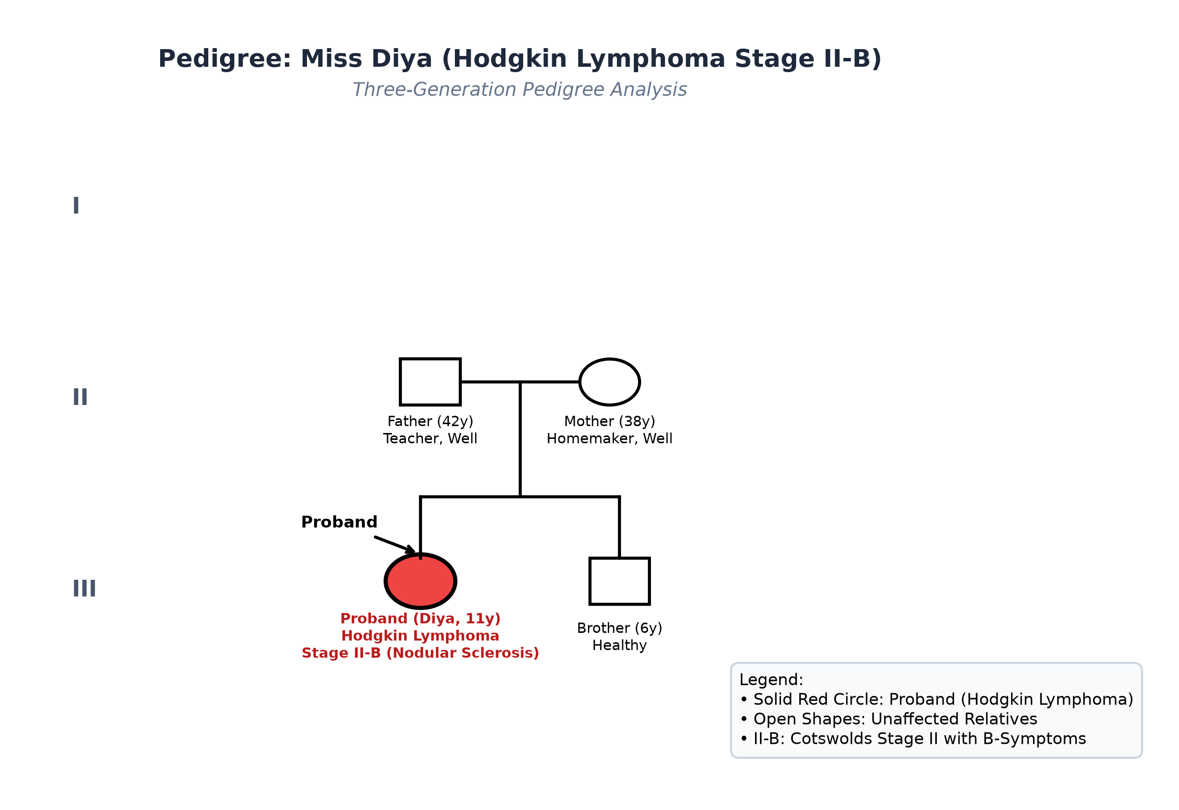
- Father 42 years, teacher, healthy; Mother 38 years, homemaker, healthy.
- Younger brother (6 years old) is attending school, healthy.
- No family history of lymphoma, leukemia, or autoimmune connective tissue disorders.
Immunization history
- Fully immunized per the National Immunization Schedule, including BCG scar present over left deltoid.
Dietary history
- Vegetarian diet; intake reduced by 30% over the past month due to systemic malaise.
| Food Item | Quantity | Calories (kcal) | Protein (g) |
|---|---|---|---|
| Cow's Milk (toned) | 300 mL | 180 | 9.6 |
| Roti (whole wheat, 2) | 60 g flour | 204 | 7.0 |
| Boiled Rice | 150 g cooked | 195 | 3.9 |
| Toor / Chana Dal | 40 g raw | 136 | 8.8 |
| Seasonal Mixed Sabzi | 1 bowl | 120 | 2.4 |
| Paneer Bhurji | 40 g | 110 | 7.2 |
| Apple | 1 medium (100 g) | 60 | 0.4 |
| Biscuits | 30 g | 120 | 2.1 |
| Total Observed Daily Intake | — | 1125 kcal | 41.4 g |
24-Hour Recall Deficit Analysis
$$ \text{Ideal Body Weight (IBW for 11 years, 50th centile WHO)} = 37.0\text{ kg} $$| Nutrient | Expected Intake (ICMR-NIN 2024 for IBW 37.0 kg) | Observed Intake | Deficit | Percentage Deficit |
|---|---|---|---|---|
| Energy (kcal) | $37.0\text{ kg} \times 52\text{ kcal/kg} = 1924\text{ kcal}$ | 1125 kcal | 799 kcal | 41.5% Deficit |
| Protein (g) | $37.0\text{ kg} \times 0.95\text{ g/kg} = 35.15\text{ g}$ | 41.4 g | Nil (Adequate) | 0% Deficit |
The expected calories and proteins should be calculated from the ideal body weight, not from current weight.
Socioeconomic and KAP
- Modified BG Prasad Socioeconomic Class II (Upper Middle Class).
- Urban pucca house with municipal treated water and sanitation.
Summary of History
Miss Diya, an 11-year-old female child, presents with an 8-week history of painless, progressive right supraclavicular and cervical lymphadenopathy, cyclical Pel-Ebstein fevers, drenching night sweats, $>10\%$ weight loss, and dry brassy cough when supine, without TB contact or peripheral cytopenic bleeding.
I would like to consider a provisional diagnosis of Lymphoma (most consistent with Hodgkin Lymphoma, Cotswolds Stage II-B), with suspected mediastinal lymph node involvement.
General head to toe examination
- General Appearance: Alert, adolescent girl, mild pallor, visible fullness in the right lower neck.
- Vitals:
- Heart Rate: 96 beats/minute, regular.
- Respiratory Rate: 20 breaths/minute, regular.
- Blood Pressure: $106/68\text{ mmHg}$ ($50^{\text{th}}$ centile, normotensive).
- Temperature: $37.1^\circ\text{C}$ ($98.8^\circ\text{F}$) axillary (afebrile interval).
- Capillary Refill Time: $<2$ seconds.
- Anthropometry:
| Parameter | Observed | Expected (50th WHO) | Z-score / Centile | Inference |
|---|---|---|---|---|
| Weight | 30.0 kg | 37.0 kg | $-1.5\text{ SD}$ | Significant weight loss (4 kg / 11.7%) |
| Pre-morbid Weight | 34.0 kg | 37.0 kg | $-0.5\text{ SD}$ | Baseline normal weight |
| Height | 144.0 cm | 144.5 cm | $50^{\text{th}}\text{ centile}$ | Normal Stature |
| Weight-for-Height | 30.0 kg for 144 cm | 36.0 kg | $-1.8\text{ SD}$ | Mild-to-moderate thinness |
| BMI | $14.5\text{ kg/m}^2$ | $17.2\text{ kg/m}^2$ | $-1.8\text{ SD}$ | Mild-to-moderate thinness |
- Lymph Node Examination:
- Right Anterior Cervical & Supraclavicular Chain:
- A cluster of enlarged lymph nodes situated along the lower third of the sternocleidomastoid muscle and right supraclavicular fossa.
- Largest mass measures $4.0\text{ cm} \times 3.0\text{ cm}$; firm-to-rubbery consistency, smooth surface, discrete, non-matted, freely mobile over underlying muscle, and completely non-tender.
- Overlying skin is normal, mobile, no erythema, warmth, ulceration, or discharging sinus.
- Other Lymph Node Basins:
- Left cervical, bilateral axillary, epitrochlear, and inguinal lymph node regions are completely free of palpable enlargement.
- Right Anterior Cervical & Supraclavicular Chain:
- Signs of Superior Mediastinal Syndrome (SMS):
- No facial plethora, periorbital edema, or chemosis.
- No engorgement of superficial veins over the neck or anterior chest wall.
- No inspiratory stridor or hoarseness; cough elicited on lying flat.
- Systemic Signs:
- Pallor: Mild conjunctival pallor.
- Icterus, Cyanosis, Clubbing, Edema: Absent.
- Skin: Normal, no excoriation marks (no active pruritus).
Systemic Examination
Respiratory System & Mediastinum
- Trachea is midline, no tracheal tug.
- Retrosternal Dullness: Percussion over the upper half of the manubrium sterni elicits retrosternal dullness extending 2 cm to the right of the sternal border $\to$ Suggests Anterior Mediastinal Mass.
- Breath sounds: Normal vesicular breath sounds bilaterally; no wheezing, rhonchi, or crepitations.
Abdomen
- Flat, soft, non-tender.
- Liver & Spleen: Neither liver nor spleen is palpable below the costal margins; liver span 10 cm, no splenic dullness.
- No abdominal masses or ascites.
Cardiovascular & Neurological Systems
- Normal heart sounds; no murmurs or pericardial rub. Nervous system examination normal; GCS 15/15.
Summary
Miss Diya, an 11-year-old female child, presents with an 8-week history of painless, firm, rubbery, discrete right cervical and supraclavicular lymphadenopathy ($4 \times 3\text{ cm}$), retromansternal percussion dullness, dry cough when supine, and prominent constitutional B-symptoms (cyclical fever, drenching night sweats, $11.7\%$ weight loss), in the absence of hepatosplenomegaly or peripheral cytopenias.
Final Clinical Diagnosis: Hodgkin Lymphoma (likely Nodular Sclerosis subtype), Cotswolds / Ann Arbor Stage II-B (Two lymph node regions involved on the same side of the diaphragm: right cervical/supraclavicular and mediastinal, with B-symptoms), without evidence of Superior Vena Cava syndrome.
Differential Diagnosis
| Disorder | Points IN FAVOR | Points AGAINST |
|---|---|---|
| Hodgkin Lymphoma (HL) | Adolescent age; rubbery, painless, discrete supraclavicular nodes; classic B-symptoms; mediastinal widening | Primary Diagnosis |
| Tuberculous Lymphadenitis | Endemic India; prolonged fever, weight loss, cervical adenopathy | TB nodes are typically matted, periadenitic, adherent, caseating, with cold abscess/sinuses; lacks Pel-Ebstein fever or supraclavicular rubbery nodes |
| Non-Hodgkin Lymphoma (NHL) | Rapidly enlarging lymph nodes, mediastinal mass | Pediatric NHL has a fulminant doubling time (days to weeks), diffuse extranodal spread, and rapidly precipitates airway collapse; HL has slower course (months) |
| Reactive Lymphadenopathy | Cervical node enlargement | Reactive nodes are small ($<1.5\text{ cm}$), tender, soft, associated with acute upper respiratory infection, and resolve in 2-4 weeks |
| Infectious Mononucleosis | Fever, cervical lymphadenopathy | Associated with acute exudative tonsillitis, splenomegaly, and atypical lymphocytes; does not persist for 8 weeks with 11% weight loss |
Investigation Protocol & Diagnostic Workup
flowchart TD
    A["Adolescent with Rubbery Supraclavicular Adenopathy & B-Symptoms"] --> B["Erect Chest Radiograph: Assess Mediastinal Mass Ratio (MMR)"]
    B --> C["EXCISIONAL Lymph Node Biopsy of Cervical Node (NOT FNAC!)"]
    C --> D["Histopathology & IHC: Identify Reed-Sternberg Cells (CD15+, CD30+, Pax-5+)"]
    D --> E["Whole-Body 18F-FDG PET-CT: Precise Metabolic Staging (Ann Arbor I to IV)"]
    E --> F["Laboratory Workup: CBC, ESR, Albumin, Ferritin, LDH, Renal/Hepatic Panels"]
    F --> G["Determine Stage: Stage II-B with Mediastinal Involvement"]
    G --> H["Initiate Risk-Adapted Chemotherapy: OEPA / ABVD Regimen"]
1. The Definitive Diagnostic Procedure (VIVA TRAP)
- EXCISIONAL Lymph Node Biopsy (MANDATORY RULE):
- Fine Needle Aspiration Cytology (FNAC) is STRONGLY CONTRAINDICATED / INSUFFICIENT to diagnose or subtype lymphoma because it aspirates only single cells, destroying the nodal architectural context and almost always missing the rare, scattered Reed-Sternberg cells (which comprise $<1-2\%$ of the tumor mass)!
- Excision of the entire intact right supraclavicular lymph node under local anesthesia is performed.
- Histopathology & Immunohistochemistry (IHC):
- Demonstrates collagen bands dividing the lymphoid tissue into circumscribed nodules (Nodular Sclerosis subtype).
- Background rich in polyclonal lymphocytes, histiocytes, plasma cells, and eosinophils.
- Identification of classical Reed-Sternberg (RS) cells: large binucleated giant cells with prominent inclusion-like eosinophilic nucleoli ('owl-eye' appearance) and lacunar cell variants.
- IHC Profile: Tumor cells are strongly positive for CD30 ($100\%$), CD15 ($90\%$), and weakly positive for Pax-5; negative for CD45 (LCA) and CD20.
2. Staging Modalities (18F-FDG PET-CT)
- Whole-Body 18F-Fluorodeoxyglucose (FDG) PET-CT:
- Intense FDG avidity in right lower cervical and supraclavicular lymph nodes (SUVmax 11.2).
- Intense FDG avidity in an Anterior Mediastinal Nodal Mass measuring $5.2 \times 4.1\text{ cm}$ (SUVmax 13.8).
- Mediastinal-to-Thoracic Ratio (MMR): $0.28$ ($<0.33$, confirms non-bulky mediastinal disease).
- Subdiaphragmatic nodes (para-aortic, mesenteric, iliac) show normal baseline metabolic activity.
- Liver and spleen are metabolically normal without focal lesions.
- Skeletal system shows no FDG-avid bone lesions.
- Bone Marrow Trephine Biopsy: Bilateral posterior iliac crest marrow biopsies show normocellular marrow negative for lymphoma infiltration.
3. Laboratory Biomarkers & Stage Grouping
- Erythrocyte Sedimentation Rate (ESR): $78\text{ mm/hr}$ (Markedly elevated; adverse prognostic marker if $>50\text{ mm/hr}$).
- Complete Blood Count: Hb $10.2\text{ g/dL}$ (mild anemia of chronic disease), TLC $11,200/\mu\text{L}$ with $8\%$ eosinophils, Platelets $410,000/\mu\text{L}$.
- Serum Albumin: $3.8\text{ g/dL}$; Serum LDH: $480\text{ U/L}$ (moderately elevated).
- Renal and Hepatic panels completely normal.
- Staging Formulation: Ann Arbor / Cotswolds Stage II-B (Two lymph node regions on the same side of diaphragm with B-symptoms).
Risk-Adapted Multimodal Management Plan
1. Chemotherapy Protocol: Pediatric Oncology Group (OEPA / COPDAC Regimen)
Modern pediatric Hodgkin lymphoma regimens minimize cumulative anthracycline and bleomycin doses to prevent long-term cardiotoxicity and pulmonary fibrosis, and eliminate alkylating agents (procarbazine) to preserve male and female fertility:
- Intermediate-Risk Protocol (Stage II-B):
- Induction: 2 Cycles of OEPA:
- O (Vincristine / Oncovin): $1.5\text{ mg/m}^2$ IV on Days 1, 8, 15.
- E (Etoposide): $100\text{ mg/m}^2$ IV on Days 1 to 5.
- P (Prednisone): $60\text{ mg/m}^2/\text{day}$ orally on Days 1 to 15.
- A (Doxorubicin / Adriamycin): $40\text{ mg/m}^2$ IV on Days 1 and 15.
- Induction: 2 Cycles of OEPA:
- Interim PET-CT (After 2 Cycles of OEPA):
- If Adequate Response (Deauville Score 1 or 2): Administer 2 Cycles of COPDAC (Cyclophosphamide, Vincristine, Prednisone, Dacarbazine) and OMIT RADIOTHERAPY COMPLETELY!
- If Inadequate Response: Administer involved-field radiotherapy (IFRT) at reduced doses ($15-20\text{ Gy}$).
2. Long-Term Survivorship & Toxicity Monitoring
- Cardiac Surveillance: Baseline and annual echocardiogram (Ejection Fraction / Strain imaging) to monitor for anthracycline-induced cardiomyopathy.
- Thyroid Surveillance: Annual TSH monitoring if mediastinal/cervical radiation is utilized.
- Secondary Malignancy Screening: Breast cancer screening via MRI/mammography starting at age 25 or 8 years post-therapy for females receiving thoracic irradiation.
- Overall Prognosis: $>90-95\%$ 5-year event-free survival with modern response-adapted protocols.