
Master Harsh, a 2.5 year old boy, 2nd order child born of a non-consanguineous marriage from Ahmedabad, Gujarat presented with complaints of bluish discoloration of lips, tongue, and fingertips since 4 months of age, episodes of severe crying followed by deepening bluishness and fast breathing (cyanotic spells) since 8 months of age, and a habit of squatting while playing since 1.5 years of age.
- Central cyanosis appearing in early infancy (worsening on exertion or crying)
- Hypoxic / Hypercyanotic / "Tet" spells (paroxysmal hyperpnea, irritability, limpness, loss of consciousness)
- Squatting posture during exertion (physiologically increases SVR, decreasing right-to-left shunt)
- Clubbing of digits and exertional fatigue without heart failure
HOPI
The history is dated back to 4 months of age when the mother first noticed bluish discoloration of the child's lips and tongue, especially during bouts of crying.
Always establish whether pulmonary blood flow is decreased (oligemic - e.g. TOF) or increased (plethoric - e.g. TGA, Truncus). In TOF, pulmonary blood flow is restricted by RV outflow tract obstruction (RVOTO); hence, infants DO NOT develop congestive cardiac failure or recurrent chest infections, but instead present with hypoxemia, spells, and polycythemia.
- Central Cyanosis:
- Noticed initially over lips and mucous membranes of the mouth at 4 months of age during crying.
- Gradually became persistent at rest by 8-10 months of age.
- Deepens markedly during crying, defecation, fever, or physical exertion.
- No history of differential cyanosis (lower limbs bluer than upper limbs, or vice versa) Differential cyanosis suggests PDA with Eisenmenger or Coarctation with right-to-left ductal flow. In TOF, cyanosis is uniform and generalized.
- Cyanotic Spells (Hypercyanotic / Anoxic / Tet Spells):
- First episode occurred at 8 months of age early in the morning after waking up.
- Child cried intensely, developed deep bluish-black discoloration of lips and face, breathed very fast and deeply (paroxysmal hyperpnea), became excessively irritable, and subsequently became limp and drowsy for 15 minutes before slowly recovering.
- Mother reports 4 such episodes over the past 6 months, typically precipitated by morning bowel movements, dehydration during diarrhea, or post-vaccination fever.
- Mother learned that holding the child in a tight chest-to-knee position helped shorten the episodes Knee-chest position kinks femoral arteries, sharply elevating systemic vascular resistance (SVR), thereby forcing deoxygenated blood across the stenotic pulmonary valve rather than through the VSD into the aorta.
- Squatting Posture:
- Since the child began walking at 16 months, the mother noted that after running or playing for 2-3 minutes, he would sit down on his haunches in a squatting position.
- After 3-5 minutes of squatting, the child would stand up and resume playing Squatting: 1) increases SVR by compressing femoral vessels; 2) increases venous return to the heart; 3) sequesters highly desaturated, acidotic blood in lower limbs.
- Absence of Heart Failure and Recurrent Pneumonia:
- No history of tachypnea at rest, suck-rest-suck feeding fatigue, forehead sweating, or recurrent pneumonia admissions Severe infundibular pulmonary stenosis protects pulmonary vasculature from volume/pressure overload, rendering pulmonary fields oligemic.
- Negative History:
- No history of focal seizures, weakness of one side of the body, or sudden altered sensorium Rules out cerebrovascular thrombosis (due to severe secondary polycythemia) and paradoxical cerebral abscess.
- No history of prolonged unexplained fever, petechiae, splinter hemorrhages, or bone pains Rules out infective endocarditis.
- No history of hemoptysis or epistaxis Rules out bleeding diathesis from thrombocytopenia / coagulopathy of chronic cyanosis, and rupture of major aortopulmonary collateral arteries (MAPCAs).
- No history of maternal lithium, retinoid exposure, or maternal phenylketonuria.
Past History
- History of hospitalization at 14 months of age for a prolonged cyanotic spell triggered by acute gastroenteritis, managed in PICU with knee-chest position, oxygen, IV morphine, IV fluids, and IV sodium bicarbonate.
- Started on oral propranolol ($1.5 ext{ mg/kg/day}$) prophylaxis post-discharge with partial reduction in spell frequency.
- No history of stroke, blood transfusion, or therapeutic phlebotomy.
Family history
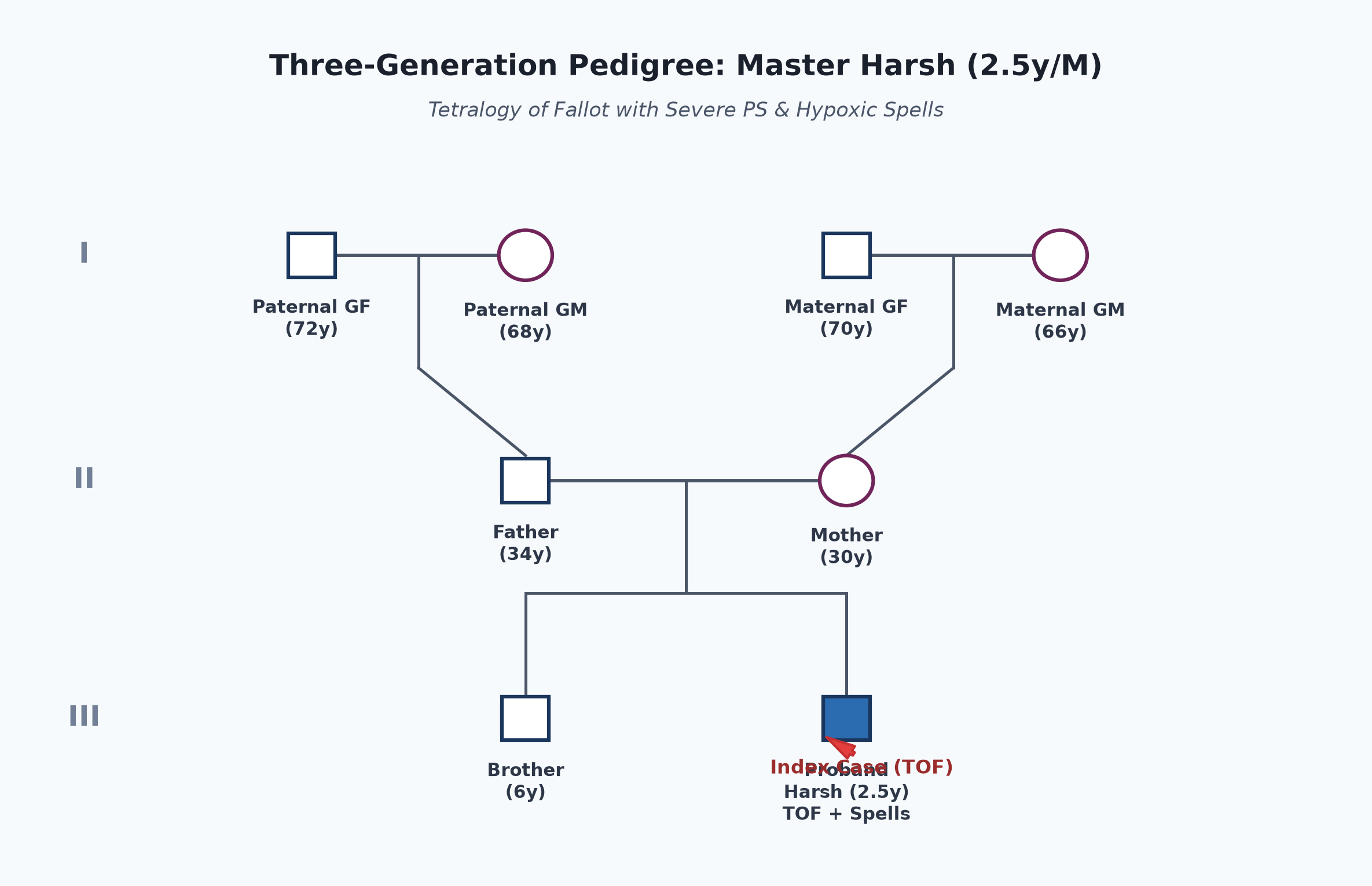
- Non-consanguineous marriage.
- Father 34 years, clerk; mother 30 years, homemaker. Both healthy.
- Elder brother (6 years) is asymptomatic, going to school, with normal cardiovascular examination.
- No family history of congenital heart disease, cleft palate, hypocalcemia, or early infant deaths.
Immunization history
- Fully immunized for age as per National Immunization Schedule (UIP): BCG, Hepatitis B, OPV, Pentavalent (1-3), Rotavirus, PCV, MR-1, and DPT/OPV booster at 16-24 months.
- Mother was appropriately counselled to administer paracetamol proactively post-vaccination to prevent fever-induced cyanotic spells.
Dietary history
- Weaned at 6 months; currently takes family diet comprising roti, dal, rice, milk, and seasonal fruits.
- Appetite is fair; does not show feeding fatigue.
| Food Item | Quantity | Calories (kcal) | Protein (g) |
|---|---|---|---|
| Cow's milk | 350 mL | 234 | 11.2 |
| Rice & Roti | 1 bowl rice + 2 rotis | 380 | 8.5 |
| Dal (thick) | 1 medium bowl | 120 | 6.8 |
| Vegetables & Fruits | 1 small cup | 75 | 1.2 |
| Total Observed Intake | — | 809 kcal | 27.7 g |
24-Hour Recall Deficit Analysis
$$ ext{Ideal Body Weight (IBW for 2.5 years, 50th centile WHO)} = 13.0 ext{ kg} $$| Nutrient | Expected Intake (ICMR-NIN 2024 for 1-3 years / 13 kg) | Observed Intake | Deficit | Percentage Deficit |
|---|---|---|---|---|
| Energy (kcal) | $1110 ext{ kcal/day}$ | 809 kcal | 301 kcal | 27.1% Deficit |
| Protein (g) | $12.5 ext{ g/day}$ | 27.7 g | 0 g | No Deficit (Excess) |
The expected calories and proteins should be calculated from the ideal body weight, not from current weight.
Socioeconomic and KAP
- Belongs to Modified BG Prasad Class III (Lower-middle socioeconomic group).
- Lives in an urban pucca flat with municipal water and sanitary toilet.
- Mother is well-trained in identifying cyanotic spells and placing the child in the knee-chest position.
Summary of History
Master Harsh, a 2.5-year-old male child, second born of non-consanguineous marriage, presented with early onset central cyanosis since 4 months of age, paroxysmal hypercyanotic spells since 8 months, and exertional squatting since 1.5 years of age, without history of congestive heart failure, recurrent respiratory infections, or focal neurological deficits.
I would like to think of a Congenital Heart Disease, Cyanotic type with Decreased Pulmonary Blood Flow (Oligemic Lung Fields), clinically characteristic of Tetralogy of Fallot (TOF) with recurrent cyanotic spells, without evidence of infective endocarditis, brain abscess, or cerebrovascular thrombosis.
General head to toe examination
- Child Behavioral State: Calm, sitting on mother's lap, cooperative (Prechtl State 3).
- Vitals:
- Heart Rate: 108 beats/minute, regular, good volume, all peripheral pulses palpable symmetrically; no radio-femoral delay.
- Respiratory Rate: 24 breaths/minute, normal abdominothoracic pattern, no retractions, no grunting.
- Blood Pressure: Right upper limb: $92/58 ext{ mmHg}$ ($50^{ ext{th}}$ centile for age/sex).
- Temperature: $37.0^\circ ext{C}$.
- Oxygen Saturation ($SpO_2$): 78% on room air in all four limbs at rest; drops to 64% on walking/crying.
- Anthropometry:
| Parameter | Observed | Expected (50th WHO) | Z-score / Centile | Inference |
|---|---|---|---|---|
| Weight | 10.6 kg | 13.0 kg | $-1.5 ext{ SD}$ | Mild Underweight |
| Height | 88 cm | 92.0 cm | $-1 ext{ to } -2 ext{ SD}$ | Mild Stunting |
| Weight-for-Height | 10.6 kg for 88 cm | 12.2 kg | $-1 ext{ to } -2 ext{ SD}$ | Mild Wasting |
| Head Circumference | 48.5 cm | 49.0 cm | Normal | Normal Head Growth |
- Head to Toe Findings:
- Central Cyanosis: Deep bluish discoloration clearly visible on the tongue, inner buccal mucosa, gingiva, lips, and nail beds.
- Clubbing: Grade III digital clubbing present on all fingers and toes (drumstick appearance with obliteration of Lovibond angle and positive Schamroth sign).
- Conjunctival Congestion: Prominent reddish engorgement of conjunctival vessels (secondary polycythemia).
- No pallor, jaundice, generalized lymphadenopathy, or peripheral edema.
- No syndromic facial features: No low-set ears, no micrognathia, no cleft palate (DiGeorge 22q11 microdeletion features absent).
Systemic Examination
Cardiovascular System
- Inspection:
- Chest shape: Symmetrical, no precordial bulge, no Harrison sulcus.
- Apical impulse: Visible in the left $4^{ ext{th}}$ intercostal space, inside the midclavicular line.
- No visible left parasternal heave or epigastric pulsations.
- Palpation:
- Apical Impulse: Palpable in the left $4^{ ext{th}}$ intercostal space in the midclavicular line; tapping in character.
- Parasternal Heave: Mild Grade 1/3 parasternal heave palpable along the left lower sternal border (right ventricular hypertrophy).
- Palpable Heart Sounds: Second heart sound palpable in the left $2^{ ext{nd}}$ intercostal space; represents $A_2$ (due to anteriorly transposed, dextroposed dilated aortic root). $P_2$ is impalpable.
- Thrill: Systolic thrill palpable at the left upper to mid sternal border ($2^{ ext{nd}}-3^{ ext{rd}}$ intercostal space) (infundibular and valvular pulmonary stenosis).
- Percussion:
- Heart borders normal size; no cardiomegaly on percussion (cor bovinum absent).
- Auscultation:
- First Heart Sound ($S_1$): Normal intensity.
- Second Heart Sound ($S_2$): Single and loud.Examiner Pearl: Nature of S2 in TOF
The single $S_2$ heard in TOF is composed almost entirely of the loud, closure sound of the dilated aortic root ($A_2$). The pulmonary component ($P_2$) is extremely soft, delayed, or completely inaudible due to the low pressure and low flow in the hypoplastic pulmonary trunk.
- Aortic Ejection Click: Audible along the left sternal border in early systole (dilated, overriding aortic root).
- Murmur:
- Grade 3/6, harsh, crescendo-decrescendo ejection systolic murmur (ESM) heard loudest at the left $2^{ ext{nd}}$ and $3^{ ext{rd}}$ intercostal spaces (pulmonary area).
- Radiation: Radiates towards the left clavicle and suprasternal notch, and poorly to the lung fields.
- Crucial Examiner Concept: The murmur in TOF originates entirely from the Pulmonary Stenosis (RVOTO), NOT from the VSD! The VSD in TOF is large, non-restrictive, and hemodynamically silent (no pressure gradient between RV and LV).
- During a cyanotic spell, infundibular spasm completely shuts off flow into the pulmonary artery, causing the murmur to become softer or disappear entirely.
other systems
- Respiratory System: Normal bilateral vesicular breath sounds; lung fields completely clear, no wheezes or crackles.
- Abdomen: Soft, non-tender; liver palpable 1 cm below right costal margin (soft, normal span); spleen not palpable; no ascites.
- Central Nervous System: Conscious, alert, active, oriented; no meningeal signs, no cranial nerve palsy, normal power and tone in all 4 limbs (no stroke or abscess).
Summary
Master Harsh, a 2.5-year-old male child, presented with central cyanosis since 4 months of age, paroxysmal hypercyanotic spells since 8 months, and exertional squatting since 1.5 years of age, without history of heart failure or stroke.
On examination, the child has central cyanosis with Grade III clubbing, resting $SpO_2$ of 78%, tapping apex beat, Grade 1 parasternal heave, a single loud $S_2$, an early systolic aortic ejection click, and a Grade 3/6 ejection systolic murmur at the left upper sternal border, with clear lung fields and no hepatomegaly.
Probable Clinical Diagnosis: Congenital Cyanotic Heart Disease with Decreased Pulmonary Blood Flow (Oligemic Lungs), anatomically consistent with Tetralogy of Fallot (Classic Pink-turned-Blue TOF with Severe Infundibular and Valvular Pulmonary Stenosis and Large Subaortic VSD), complicated by Recurrent Hypercyanotic Spells, without clinical evidence of infective endocarditis, polycythemia-induced thromboembolism, or cerebral abscess.
Differential Diagnosis
| Disease | Points IN FAVOR | Points AGAINST |
|---|---|---|
| Tetralogy of Fallot (TOF) | • Cyanosis appearing in early infancy • Hypercyanotic spells + squatting posture • Single loud $S_2$ + harsh ESM at pulmonary area • Clear chest, no cardiomegaly, no CCF | Primary Diagnosis |
| Pulmonary Atresia with VSD (Extreme TOF) | • Cyanotic from early infancy • Single loud $S_2$, right ventricular hypertrophy | • Cyanosis presents on day 1 with ductal closure • No ejection systolic murmur (flow is via PDA or MAPCAs generating continuous murmur) |
| Tricuspid Atresia with PS | • Cyanosis with oligemic lung fields • Single $S_2$, systolic murmur | • Left axis deviation ($-30^\circ ext{ to } -90^\circ$) and LV hypertrophy on ECG (TOF has RAD and RVH) • Prominent 'a' wave in JVP and left ventricular impulse |
| Transposition of Great Arteries (TGA) with VSD & PS | • Cyanosis, spells, single $S_2$, systolic murmur | • Cyanosis typically appears immediately after birth ($<48 ext{ hours}$) • Clinically indistinguishable without echocardiography |
Investigation Protocol & Diagnostic Workup
flowchart TD
    A["Suspected Cyanotic Heart Disease (TOF)"] --> B["1. Non-Invasive Diagnostic Workup"]
    B --> C["Chest X-Ray (PA View)"]
    B --> D["12-Lead Electrocardiogram (ECG)"]
    B --> E["Hyperoxia Test (100% O2 Challenge)"]

    A --> F["2. Confirmatory 2D Echocardiography"]
    F --> G["Anatomy of Subaortic VSD & Overriding Aorta"]
    F --> H["Severity & Anatomy of RVOTO (Infundibular vs Valvular)"]
    F --> I["Pulmonary Artery Anatomy (Nakata Index & McGoon Ratio)"]
    F --> J["Coronary Artery Anatomy (Rule out LAD crossing RVOT)"]

    A --> K["3. Hematology & Secondary Complication Surveillance"]
    K --> L["CBC: Hb, Hct, RBC Indices (Microcytic Hypochromic Polycythemia)"]
    K --> M["Coagulation Screen: PT, INR, aPTT, Fibrinogen"]
1. Chest Radiography (CXR - Posteroanterior View)
- Classic "Cœur en sabot" (Boot-shaped heart):
- Cardiac size is normal (no cardiomegaly, $CTR < 0.50$).
- Upturned, rounded apex elevated above the diaphragm (due to right ventricular concentric hypertrophy).
- Conspicuous concavity in the pulmonary bay / waist (due to hypoplastic main pulmonary artery).
- Pulmonary Oligemia: Markedly attenuated pulmonary vascular markings throughout both lung fields (black, hyperlucent lung fields).
- Right-Sided Aortic Arch: Visible in approximately 25% of TOF cases (indenting the right side of the trachea).
2. Electrocardiogram (12-Lead ECG)
- Right Axis Deviation: QRS frontal axis $+120^\circ ext{ to } +150^\circ$.
- Right Ventricular Hypertrophy (RVH):
- Tall R wave in lead V1 ($R > 15 ext{ mm}$), upright T wave in V1 after 7 days of life.
- Deep S wave in lead V6 ($R/S ext{ ratio} < 1$).
- Sudden transition ("voltage drop-off") between V1 and V2 without biventricular hypertrophy.
- Right Atrial Enlargement (P pulmonale): Peaked P waves $> 2.5 ext{ mm}$ in lead II.
3. Hyperoxia Test (100% Oxygen Challenge)
- Administer 100% normobaric oxygen via tight-fitting mask with reservoir bag for 10 minutes:
- In parenchymal pulmonary disease: $PaO_2$ rises $> 150-200 ext{ mmHg}$.
- In fixed intracardiac right-to-left shunt (TOF): $PaO_2$ remains $< 100 ext{ mmHg}$ and $SpO_2$ shows minimal or no change ($<5\%$).
4. 2D-Echocardiography with Color Doppler (Definitive Modality)
- Subaortic VSD: Alignment and dimension of large malalignment VSD.
- Aortic Overriding: Degree of aortic root dextroposition overriding the ventricular septum ($<50\%$).
- RVOTO Assessment: Continuous-wave Doppler measuring peak systolic gradient across RVOT ($>60 ext{ mmHg}$ indicates severe obstruction).
- Branch Pulmonary Artery Sizing: Sizing RPA and LPA diameters to calculate: $$ ext{McGoon Ratio} = rac{ ext{Diameter of RPA} + ext{Diameter of LPA}}{ ext{Diameter of Descending Aorta at Diaphragm}} \quad ( ext{Normal } \ge 2.0; \ge 1.2 ext{ indicates suitability for repair}) $$ $$ ext{Nakata Index} = rac{ ext{Cross-sectional Area of RPA} + ext{LPA}}{ ext{Body Surface Area (BSA)}} \quad (\ge 150 ext{ mm}^2/ ext{m}^2 ext{ indicates adequate pulmonary bed}) $$
- Coronary Artery Origin: Exclude anomalous origin of Left Anterior Descending (LAD) coronary artery arising from the Right Coronary Artery crossing the RVOT (precludes standard RVOT transannular incisional patching).
5. Complete Blood Count & Coagulation Profile
- Polycythemia: $Hb = 17.8 ext{ g/dL}$, $Hematocrit (Hct) = 56\%$.
- Relative Iron Deficiency Alert: MCV 68 fL, MCH 22 pg. Critical DNB Point: Even with high Hb, microcytic hypochromic red cells are rigid and significantly increase blood viscosity, exponentially elevating stroke risk. Supplemental iron must be given!
Management Plan
1. Acute Management of a Cyanotic (Tet) Spell
The acute hypercyanotic spell is a medical emergency requiring rapid sequential intervention:
- Step 1: Knee-Chest Position: Flex knees firmly onto the chest. Calms the child and immediately elevates Systemic Vascular Resistance (SVR), decreasing right-to-left shunting.
- Step 2: High-Flow Oxygen: Administer 100% oxygen via tight face mask (vasodilates pulmonary bed).
- Step 3: Morphine Sulfate: $0.1-0.2 ext{ mg/kg}$ subcutaneous or intramuscular (or IV slowly). Depresses respiratory center drive, terminates hyperpnea, relieves infundibular spasm, and allays anxiety.
- Step 4: Intravenous Fluid Bolus: Normal Saline $10-20 ext{ mL/kg}$ rapid IV bolus to expand intravascular volume and optimize RV preload.
- Step 5: Sodium Bicarbonate: $1-2 ext{ mEq/kg}$ IV diluted 1:1 with distilled water over 5 minutes to correct metabolic lactic acidosis (acidosis is a potent pulmonary vasoconstrictor).
- Step 6: Beta-Blocker (IV Esmolol or Propranolol): IV Esmolol ($500 ext{ mcg/kg}$ load over 1 min followed by $50 ext{ mcg/kg/min}$) or IV Propranolol ($0.05-0.1 ext{ mg/kg}$ slow IV) to relax infundibular muscular spasm.
- Step 7: Vasopressor (Phenylephrine): $5-10 ext{ mcg/kg}$ IV bolus; pure alpha-1 agonist that sharply raises SVR, reversing the shunt back into the pulmonary circulation.
2. Prophylactic Medical Therapy (Bridging to Surgery)
- Oral Propranolol: $1-3 ext{ mg/kg/day}$ divided every 6 to 8 hours. Prevents catecholamine-mediated infundibular muscle spasm, significantly reducing spell recurrence.
- Oral Iron Therapy: Elemental iron $3 ext{ mg/kg/day}$ for 3 months to treat relative iron deficiency and improve red cell deformability, even in the presence of elevated hemoglobin.
- Hydration: Avoid dehydration at all times; prompt oral or IV rehydration during diarrheal or febrile illnesses to prevent hemoconcentration and sagittal sinus thrombosis.
3. Definitive Surgical Correction
- Optimal Age: Elective complete intracardiac repair is performed between 3 to 12 months of age in modern pediatric cardiac surgery centers.
- Complete Intracardiac Repair (Primary Open Heart Surgery):
- Median sternotomy under cardiopulmonary bypass with cardioplegic arrest.
- Patch closure of the non-restrictive subaortic VSD (rerouting LV flow strictly to the aorta).
- Resection of obstructing infundibular muscular bands (infundibulectomy).
- Relief of RVOTO: Pulmonary valvotomy; pulmonary artery patch augmentation; or transannular pericardial patch (TAP) if the pulmonary valve annulus $Z ext{-score} < -2.5$.
- Palliative Shunt (Modified Blalock-Taussig-Thomas Shunt [mBTT]):
- Interposition Gore-Tex tube graft between the subclavian artery and the ipsilateral branch pulmonary artery.
- Reserved for: Severe refractory spells in small neonates ($<3 ext{ kg}$), hypoplastic pulmonary arteries ($McGoon < 1.2$, $Nakata < 150 ext{ mm}^2/ ext{m}^2$), or anomalous crossing LAD coronary artery.