
Master Siddharth, a 6 month old boy, 2nd order child born of a non-consanguineous marriage from Madurai, Tamil Nadu presented with complaints of difficulty in breathing and fast breathing since 2 months of age, feeding difficulty with excessive forehead sweating since 2.5 months of age, and failure to gain weight.
- Fast breathing, chest indrawing, and recurrent lower respiratory tract infections (increased pulmonary blood flow)
- Feeding fatigue ("suck-rest-suck" cycle) taking >30 minutes per feed
- Profuse forehead sweating during feeds (compensatory sympathetic overdrive)
- Poor weight gain and growth faltering (hypermetabolic state + inadequate caloric intake)
HOPI
The history is dated back to 2 months of age when the mother first noticed fast breathing in the child while at rest.
Always establish the exact age of symptom onset. High pulmonary vascular resistance (PVR) in the immediate neonatal period limits left-to-right shunting. As physiological fall in PVR occurs between 4 to 8 weeks, left-to-right shunt surges, precipitating congestive cardiac failure and hyperdynamic pulmonary arterial hypertension (PAH).
- Difficulty in Breathing / Fast Breathing:
- Noticed since 2 months of age, insidious in onset and gradually progressive.
- Present throughout the day, persistent even during sleep.
- Associated with lower chest wall indrawing (subcostal and intercostal retractions) Points to decreased lung compliance from pulmonary venous and capillary congestion.
- Exacerbated during feeding and crying; no positional relief.
- Feeding Difficulties (Infantile Heart Failure):
- Mother noticed that the baby was unable to breastfeed continuously since 2.5 months of age.
- Baby latches on, sucks for 1-2 minutes, leaves the breast breathless and exhausted, rests for 3-5 minutes, and sucks again (suck-rest-suck cycle).
- Total feeding duration exceeded 40-45 minutes per feed with baby falling asleep exhausted without satiety.
- Associated with excessive cold diaphoresis exclusively over the forehead and scalp during feeds Reflects maximal adrenergic sympathetic discharge to maintain stroke volume during feeding exertion.
- Failure to Gain Weight (Growth Faltering):
- Birth weight was 2.9 kg. Current weight at 6 months is 4.8 kg (expected ~7.5 kg).
- Mother noted loose clothes and prominent ribs over the past 3 months despite frequent nursing attempts.
- Recurrent Lower Respiratory Infections:
- History of two episodes of fever, worsening fast breathing, and cough requiring hospitalization at 3 months and 5 months of age, treated with intravenous antibiotics and nebulization Hyperdynamic pulmonary circulation with engorged pulmonary vessels compresses bronchioles, impairing mucus clearance and predisposing to recurrent infections.
- Negative History:
- No history of bluish discoloration of lips, tongue, oral mucosa, or nail beds at rest or during crying Rules out cyanotic congenital heart disease and shunt reversal (Eisenmenger syndrome).
- No history of paroxysmal hyperpnea, severe irritability followed by limpness, or squatting equivalents Rules out hypoxic/cyanotic spells typical of Tetralogy of Fallot.
- No history of prolonged unexplained fever, petechial rashes, hematuria, or painful skin lesions Rules out infective endocarditis.
- No history of facial puffiness, abdominal distension, or pedal swelling Rules out predominant right-sided systemic venous congestion.
- No history of stridor, hoarseness of cry, or dysphagia Rules out vascular ring compressing trachea/esophagus and Ortner syndrome (left recurrent laryngeal nerve compression by dilated pulmonary artery).
- No history of maternal gestational diabetes, teratogenic drug intake (lithium, phenytoin), or rash in the first trimester Rules out syndromic/teratogenic cardiac embryopathy.
Past History
- History of two hospital admissions for severe bronchopneumonia with heart failure at 3 and 5 months of age, managed with oxygen, IV furosemide, and oral enalapril.
- No history of neonatal seizures, jaundice requiring phototherapy, or prior cardiac catheterization.
Family history
- Born of second-degree consanguineous marriage (parents are maternal first cousins).
- Father 32 years, factory employee; mother 28 years, homemaker. Both healthy.
- Elder sibling (female, 3.5 years) is asymptomatic with normal growth and no murmur.
- No family history of sudden cardiac death, congenital heart defects, cardiomyopathy, or early pacemaker implantation.
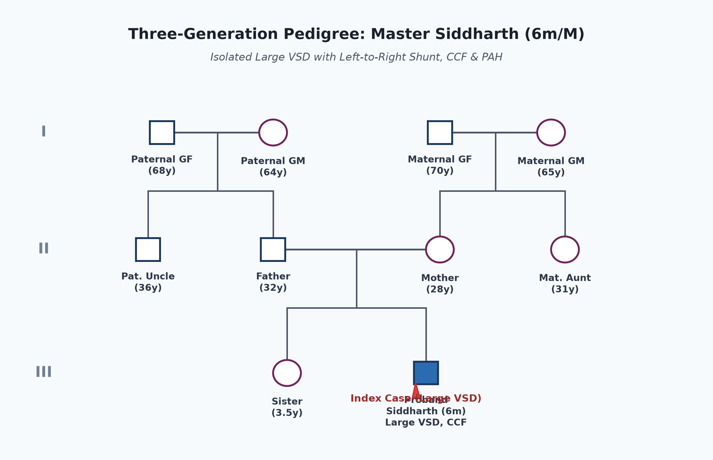
Immunization history
- Received BCG, OPV-0, and Hepatitis B at birth.
- Received Pentavalent (1, 2, 3), Rotavirus, PCV, and fIPV at 6, 10, and 14 weeks as per the National Immunization Schedule (UIP).
- Optional vaccines: Annual Influenza and Hepatitis A not yet administered.
Dietary history
- Exclusively breastfed till 5.5 months of age.
- Thin rice gruel and diluted cow's milk introduced 2 weeks ago (inadequate complementary feeding).
- Mother feeds 6-7 times in 24 hours, but effective intake is limited due to suck-rest-suck fatigue.
| Food Item | Quantity | Calories (kcal) | Protein (g) |
|---|---|---|---|
| Breast milk | ~400 mL effective | 268 | 4.4 |
| Rice gruel (watery) | 50 mL | 25 | 0.4 |
| Diluted cow's milk (1:1) | 100 mL | 33 | 1.6 |
| Total Observed Intake | — | 326 kcal | 6.4 g |
24-Hour Recall Deficit Analysis
$$ ext{Ideal Body Weight (IBW for 6 months, 50th centile WHO)} = 7.9 ext{ kg} $$| Nutrient | Expected Intake (ICMR-NIN 2024 for IBW 7.9 kg) | Observed Intake | Deficit | Percentage Deficit |
|---|---|---|---|---|
| Energy (kcal) | $7.9 ext{ kg} imes 90 ext{ kcal/kg} = 711 ext{ kcal}$ | 326 kcal | 385 kcal | 54.1% Deficit |
| Protein (g) | $7.9 ext{ kg} imes 1.25 ext{ g/kg} = 9.88 ext{ g}$ | 6.40 g | 3.48 g | 35.2% Deficit |
The expected calories and proteins should be calculated from the ideal body weight, not from current weight.
Socioeconomic and KAP
- Belongs to Modified BG Prasad Socioeconomic Class III (Middle Class).
- Urban-rural semi-pucca house with municipal tap water supply and adequate ventilation.
- Parents understand that the child has a "hole in the heart" requiring specialized surgical repair, but were hesitant due to financial constraints and fear of surgery.
Summary of History
Master Siddharth, a 6-month-old male infant, 2nd order child born of consanguineous parentage, presented with persistent tachypnea, feeding fatigue with suck-rest-suck pattern, forehead diaphoresis, failure to thrive, and recurrent lower respiratory tract infections since 2 months of age, without history of central cyanosis, cyanotic spells, or infective endocarditis features.
I would like to think of a Congenital Heart Disease, Acyanotic type, with a Left-to-Right Shunt (likely Large Ventricular Septal Defect or Patent Ductus Arteriosus), complicated by Congestive Cardiac Failure and Hyperdynamic Pulmonary Arterial Hypertension, with severe acute malnutrition.
General head to toe examination
- Child Behavioral State: Examined in quiet wakefulness (Prechtl State 3), resting comfortably on mother's lap in propped-up position, non-crying.
- Vitals:
- Heart Rate: 154 beats/minute (tachycardia), regular, normal volume, all peripheral pulses (radial, brachial, femoral, dorsalis pedis) palpable bilaterally; no radio-femoral delay, no bounding pulses.
- Respiratory Rate: 62 breaths/minute (tachypnea), thoracoabdominal, with prominent subcostal and intercostal retractions and mild alar flaring.
- Blood Pressure: Right upper limb (supine, appropriate cuff): $82/48 ext{ mmHg}$ ($50^{ ext{th}}$ centile for age/sex).
- Temperature: $36.8^\circ ext{C}$ (afebrile).
- Oxygen Saturation ($SpO_2$): 96% on room air in right upper limb and both lower limbs.
- Anthropometry:
| Parameter | Observed | Expected (50th WHO) | Z-score / Centile | Inference |
|---|---|---|---|---|
| Weight | 4.8 kg | 7.9 kg | $< -3 ext{ SD}$ | Severe Underweight |
| Length | 63 cm | 67.6 cm | $-2 ext{ to } -3 ext{ SD}$ | Moderate Stunting |
| Weight-for-Length | 4.8 kg for 63 cm | 6.8 kg | $< -3 ext{ SD}$ | Severe Acute Malnutrition (Wasting) |
| Head Circumference | 41.5 cm | 43.3 cm | $-1 ext{ to } -2 ext{ SD}$ | Normal / Sparing |
| Mid-Upper Arm Circumference | 11.2 cm | >12.5 cm | $< 11.5 ext{ cm}$ | Severe Wasting |
- Head to Toe Findings:
- Pallor present (mild conjunctival and palmar).
- No central or peripheral cyanosis, no clubbing, no pedal edema.
- Dysmorphic features: No epicanthic folds, no flat facial profile, no low-set ears (Down syndrome features absent).
- BCG scar present over left deltoid (4 mm).
Systemic Examination
Cardiovascular System
- Inspection:
- Chest shape: Precordial bulge present over the left parasternal region with visible hyperdynamic cardiac pulsations.
- Apical impulse: Visible in the left $5^{ ext{th}}$ intercostal space, $1 ext{ cm}$ lateral to the midclavicular line.
- Epigastric pulsations and left parasternal pulsations visible.
- No dilated superficial veins or surgical scars.
- Palpation:
- Apical Impulse: Palpable in the left $5^{ ext{th}}$ intercostal space, $1 ext{ cm}$ lateral to the midclavicular line; hyperdynamic in character (volume overload of left ventricle).
- Parasternal Heave: Grade 2/3 left parasternal heave present along the left $3^{ ext{rd}}$ and $4^{ ext{th}}$ intercostal spaces (right ventricular hypertrophy from PAH).
- Palpable Heart Sounds: Palpable second heart sound ($P_2$) in the left $2^{ ext{nd}}$ intercostal space (pulmonary area).
- Thrill: Systolic thrill palpable along the left lower sternal border ($3^{ ext{rd}}-4^{ ext{th}}$ intercostal space), best felt in expiration with the child sitting forward.
- Percussion:
- Right heart border percussed along the right parasternal line in the $4^{ ext{th}}$ intercostal space (right atrial dilatation).
- Left heart border percussed outside the midclavicular line in the $5^{ ext{th}}$ intercostal space.
- Upper border of liver dullness percussed in the right $5^{ ext{th}}$ intercostal space.
- Auscultation:
- First Heart Sound ($S_1$): Normal intensity, best heard at the apex.
- Second Heart Sound ($S_2$): Split is narrow but physiological; pulmonary component ($P_2$) is loud and accentuated, clearly heard at the apex and louder than $A_2$ at the base (indicates Pulmonary Arterial Hypertension).
- Third Heart Sound ($S_3$): Present at apex (reflects rapid diastolic ventricular filling).
- Murmur 1 (Regurgitant Ventricular Shunt):
- Grade 3/6, harsh pansystolic (holosystolic) murmur heard best at the left lower sternal border ($3^{ ext{rd}}$ and $4^{ ext{th}}$ intercostal space).
- Radiation: Radiates widely across the precordium; does not radiate to the axilla or back.
- Murmur 2 (Relative Mitral Stenosis / Carey-Coombs type flow rumble):
- Soft, low-pitched mid-diastolic rumble (Grade 2/6) heard best at the cardiac apex using the bell of the stethoscope in left lateral position (indicates large left-to-right shunt with $Q_p : Q_s > 2 : 1$ generating torrent flow across the normal mitral valve orifice).
Modified Ross Heart Failure Score
| Clinical Parameter | Finding in Patient | Ross Score Assigned |
|---|---|---|
| Feeding History | Takes >40 min, suck-rest-suck cycle | 2 |
| Respiratory Rate | 62 breaths/min ($>60 ext{ bpm}$) | 2 |
| Respiratory Pattern | Marked subcostal & intercostal retractions | 2 |
| Heart Rate | 154 beats/min ($>150 ext{ bpm}$) | 2 |
| Hepatomegaly | Liver 3.5 cm palpable below right costal margin | 2 |
| Peripheral Perfusion | Warm peripheries, CRT < 2 seconds | 0 |
| Total Modified Ross Score | — | 10 / 12 (Severe Heart Failure) |
Score Interpretation: 0–2: No HF; 3–6: Mild HF; 7–9: Moderate HF; 10–12: Severe HF.
other systems
- Respiratory System:
- Tachypnea with bilateral subcostal and intercostal retractions.
- Auscultation: Bilateral symmetrical vesicular breath sounds; coarse transmission crackles and fine bibasilar crepitations heard bilaterally.
- Abdomen:
- Soft, non-distended; no superficial venous collaterals.
- Liver: Palpable 3.5 cm below the right costal margin in the midclavicular line; span 7.5 cm; soft, smooth surface, tender, round margin (congestive hepatomegaly).
- Spleen: Not palpable; no free fluid (no shifting dullness).
- Central Nervous System:
- Conscious, alert, irritable on handling; anterior fontanelle normotensive (flat, soft).
- Cranial nerves intact; normal neonatal and infant developmental reflexes for 6 months.
Summary
Master Siddharth, a 6-month-old male infant, second born of second-degree consanguineous parentage, presented with history of insidious, progressive tachypnea, suck-rest-suck feeding fatigue, forehead diaphoresis, failure to thrive, and recurrent lower respiratory tract infections since 2 months of age, without history of central cyanosis or infective endocarditis.
On examination, the infant has severe wasting ($<-3 ext{ SD}$ weight-for-length), resting tachycardia ($154 ext{ bpm}$), tachypnea ($62 ext{ cpm}$) with retractions, hyperdynamic precordium, left parasternal heave (grade 2/3), palpable thrill and loud $P_2$, a Grade 3/6 harsh pansystolic murmur at the left lower sternal border, and a Grade 2/6 mid-diastolic flow rumble at the apex, associated with congestive hepatomegaly ($3.5 ext{ cm}$) and a Modified Ross Score of 10/12.
Probable Clinical Diagnosis: Congenital Acyanotic Heart Disease with a Left-to-Right Shunt, anatomically consistent with a Large Perimembranous Ventricular Septal Defect (VSD), complicated by Severe Congestive Cardiac Failure (Modified Ross Class IV / Score 10), Hyperdynamic Pulmonary Arterial Hypertension (PAH), without clinical evidence of shunt reversal (Eisenmenger syndrome) or infective endocarditis, in a background of Severe Acute Malnutrition (SAM without edema).
Differential Diagnosis
| Disease | Points IN FAVOR | Points AGAINST |
|---|---|---|
| Large Perimembranous VSD | • Onset at 2 months (physiological PVR drop) • Precordial bulge, systolic thrill at LLSB • Harsh holosystolic murmur at left 4th ICS • Apical mid-diastolic flow murmur ($Q_p:Q_s > 2:1$) • Loud $P_2$ + parasternal heave (PAH) | Primary Diagnosis |
| Patent Ductus Arteriosus (PDA) | • Left-to-right shunt physiology • CCF, FTT, recurrent LRTI • Wide pulse pressure signs | • Pulses are normal volume (not collapsing / bounding) • Murmur is pansystolic at LLSB, not continuous "machinery" murmur at left 1st/2nd infraclavicular space |
| Ostium Secundum ASD | • Acyanotic CHD with left-to-right shunt • Precordial hyperactivity + parasternal heave | • Large ASD rarely causes severe CCF and FTT at 6 months • Murmur is pansystolic with thrill (ASD has soft ESM at pulmonary area with fixed wide split $S_2$) |
| Atrioventricular Septal Defect (AVSD) | • Early severe heart failure & marked PAH • Common in consanguinity / Down syndrome | • No syndromic stigmata of Down syndrome • ECG typically shows superior axis ($-30^\circ ext{ to } -90^\circ$); murmur lacks predominant AV valve regurgitant radiation |
Investigation Protocol & Diagnostic Workup
flowchart TD
    A["Infant with Left-to-Right Shunt & CCF (Master Siddharth)"] --> B["1. Non-Invasive Bedside Tests"]
    B --> C["Chest X-Ray (CXR PA)"]
    B --> D["12-Lead Electrocardiogram (ECG)"]
    B --> E["Pulse Oximetry (Pre & Post-ductal)"]

    A --> F["2. Definitive Confirmatory Imaging"]
    F --> G["Transthoracic Echocardiography (TTE) + Color Doppler"]
    G --> H["Define VSD Location, Size, Shunt Direction & Peak Gradient"]
    G --> I["Estimate RVSP / PASP via TR Jet Velocity"]
    G --> J["Assess LA/LV Dilatation and LVEF"]

    A --> K["3. Laboratory & Metabolic Profile"]
    K --> L["CBC, Serum Electrolytes, Renal Function, NT-proBNP"]
    K --> M["ABG: Assess Acid-Base Status & PaO2"]
1. Chest Radiography (CXR - Anteroposterior View)
- Cardiomegaly: Cardiothoracic ratio (CTR) $> 0.60$ with left ventricular apex down and out.
- Pulmonary Plethora: Increased pulmonary vascular markings extending into the outer third of lung fields; dilated main pulmonary artery segment.
- Left Atrial Enlargement: Double cardiac density, splaying of the carina ($>90^\circ$).
2. Electrocardiogram (12-Lead ECG)
- Biventricular Hypertrophy (Katz-Wachtel Phenomenon): Large equiphasic biphasic QRS complexes ($\ge 50 ext{ mm}$) in mid-precordial leads (V2, V3, V4).
- Left Ventricular Volume Overload: Deep Q waves ($>2 ext{ mm}$) and tall R waves in leads V5 and V6 with upright T waves.
- Right Ventricular Hypertrophy (PAH): Tall R wave in V1 with $R/S ext{ ratio} > 1$, right axis deviation ($+120^\circ$).
3. Transthoracic 2D-Echocardiography with Color Flow Doppler (Gold Standard)
- Defect Localization: Direct visualization of perimembranous defect in parasternal long-axis (PLAX) and short-axis (PSAX) views; measuring defect diameter against aortic valve annulus ($>50\%$ aortic annulus confirms large non-restrictive VSD).
- Shunt Hemodynamics: High-velocity left-to-right flow; low peak systolic Doppler gradient ($<30 ext{ mmHg}$) indicates equalization of RV and LV pressures (severe PAH).
- Pulmonary Artery Systolic Pressure (PASP): Derived from tricuspid regurgitation (TR) jet velocity via modified Bernoulli equation: $$ ext{RVSP} = 4 imes (V_{ ext{TR}})^2 + ext{RAP} $$
- Chamber Dimensions: Dilated left atrium and left ventricle ($Z ext{-score} > +2$); preserved left ventricular ejection fraction ($LVEF \ge 60\%$).
4. Baseline Blood & Metabolic Investigations
- CBC: Mild microcytic hypochromic anemia ($Hb = 10.2 ext{ g/dL}$); total leukocyte count normal ($8,400/\mu ext{L}$).
- Serum Electrolytes: Serum $Na^+ = 136 ext{ mEq/L}$, $K^+ = 4.2 ext{ mEq/L}$, Serum Creatinine $= 0.3 ext{ mg/dL}$ (baseline check prior to ACE inhibitor/diuretic therapy).
- Biomarker: NT-proBNP elevated ($>1500 ext{ pg/mL}$), correlating with the degree of myocardial wall stretch.
Management Plan
1. Medical Stabilization of Heart Failure
- Hospitalization: Admit to pediatric cardiology ward; nurse in semi-fowler propped-up position ($30^\circ$).
- Diuretic Therapy (Preload Reduction):
- Oral Furosemide: $1 ext{ mg/kg/dose}$ PO twice daily (titrate up to $2 ext{ mg/kg/dose}$ as needed).
- Oral Spironolactone: $1 ext{ mg/kg/dose}$ PO once daily (potassium-sparing aldosterone antagonist to prevent hypokalemic alkalosis and attenuate myocardial remodeling).
- Afterload Reduction:
- Oral Enalapril: Start low at $0.1 ext{ mg/kg/day}$ in two divided doses; titrate gradually under blood pressure and serum potassium/creatinine monitoring to $0.3-0.5 ext{ mg/kg/day}$. Reduces systemic vascular resistance, favoring systemic forward output and reducing left-to-right shunt fraction.
- Oxygen Caution: Keep room air or low-flow $O_2$ only if $SpO_2 < 90\%$. Avoid excessive high-concentration $O_2$ because oxygen is a potent pulmonary vasodilator that lowers PVR, dramatically increasing left-to-right shunt and worsening pulmonary edema.
2. Nutritional Rehabilitation in Infant Cardiac Failure
- Target Caloric Intake: High caloric requirement ($120-150 ext{ kcal/kg/day}$) and high protein ($2.5-3 ext{ g/kg/day}$) required to overcome hypermetabolic stress.
- Feeding Strategy:
- Expressed Breast Milk (EBM) fortified with Medium Chain Triglyceride (MCT) oil (1 mL per 100 mL milk) or commercial milk fortifier to reach $0.8-1.0 ext{ kcal/mL}$.
- Small, frequent feeds (every 2 to 2.5 hours) to prevent exhaustion.
- If suck-rest-suck feeding exceeds 30 minutes, switch to orogastric/nasogastric tube feeding to conserve energy expenditure.
3. Definitive Surgical Correction
- Timing of Surgery: Early definitive surgical closure is indicated before 6-9 months of age in infants with large non-restrictive VSD, intractable failure to thrive, and hyperdynamic PAH to prevent irreversible pulmonary vascular remodeling (Plexogenic Pulmonary Arteriopathy / Heath-Edwards Stage III/IV).
- Procedure: Open heart surgery under cardiopulmonary bypass with transatrial patch closure (Dacron or glutaraldehyde-treated autologous pericardial patch) via right atriotomy through the tricuspid valve orifice, meticulously avoiding the postero-inferior border to prevent surgical complete heart block (Bundle of His injury).
- Pulmonary Artery Banding (PAB): Reserved only as a temporizing palliative option if severe intractable sepsis, multiple apical "Swiss-cheese" muscular VSDs, or extreme low birth weight prohibit primary open repair.
4. Preventive Strategies & Long-Term Follow-up
- Infective Endocarditis Prophylaxis: Maintain rigorous dental hygiene. As per AHA/ACC guidelines, antibiotic prophylaxis is indicated for 6 months post-surgical patch repair, or indefinitely if there is a residual shunt adjacent to prosthetic material.
- Immunization Schedule: Complete all routine UIP immunizations; administer annual trivalent inactivated influenza vaccine and pneumococcal conjugate vaccine (PCV13).